Copyright
©The Author(s) 2018.
World J Gastroenterol. May 21, 2018; 24(19): 2130-2136
Published online May 21, 2018. doi: 10.3748/wjg.v24.i19.2130
Published online May 21, 2018. doi: 10.3748/wjg.v24.i19.2130
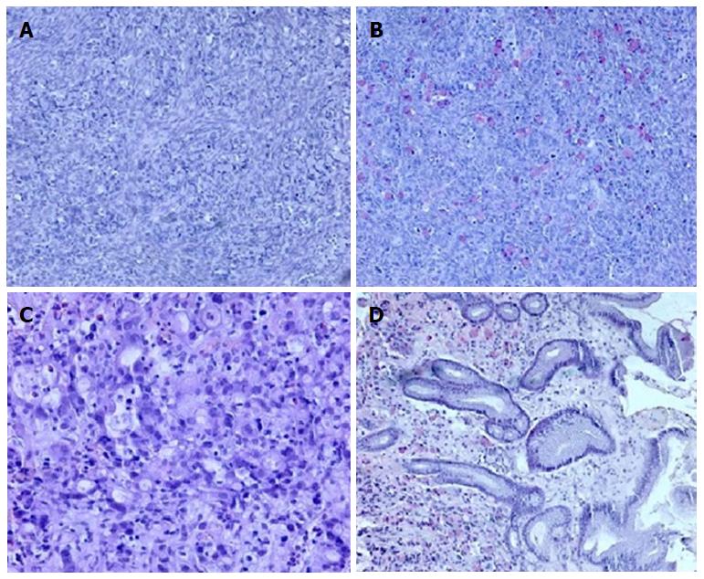
Figure 1 Histological ovary assay.
A: Signet ring of tumor cell infiltration within the ovarian stroma (10 ×, HE); B: Mucicarmine staining highlights the presence of mucin in the cytoplasm (10 ×). Histopathologic evaluation of antral mucosa showing (C) poorly differentiated carcinoma infiltration with signet ring cells (20 ×, HE) and (D) mucicarmine staining of mucin-positive cells in the gastric mucosa (10 ×).
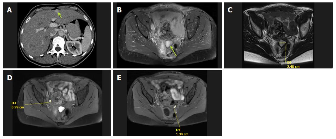
Figure 2 Abdominal computed tomography and abdominopelvic magnetic resonance scans.
A: Post-contrast computed tomography image in the portal venous phase showing a hyperintense enhancing lesion in segment V (isodense in the native phase) of the liver, which was diagnosed as a superficially located suspicious metastatic lesion; B-E: Abdominopelvic magnetic resonance scans with evident masses and suspicious nodules.
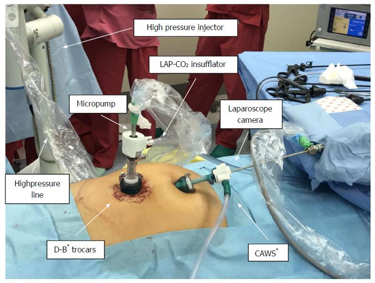
Figure 3 Schematic presentation of the pressurized intraperitoneal aerosol chemotherapy procedure.
The procedure is performed under general anesthesia and based on standard diagnostic laparoscopy procedures. Two small incisions are always made to obtain surgical access using two double-balloon secured trocars (D-B* - double-balloon secured trocars). The first one is used for the laparoscopic camera and is connected to a closed aerosol waste system (CAWS*). The second one connected to the CO2 insufflator is for the micropump nebulizer used for delivering chemotherapy under pressure via a high-pressure line.
Figure 4 Palliative open D2 gastrectomy combined with liver metastasectomy.
A: Liver metastasectomy procedure involving removal of the metastatic lesion combined with parenchyma coagulation; B: Open gastrectomy procedure showing the staple line after resection; C: Creation of a Roux-en-Y anastomosis (RNY) using sutures.
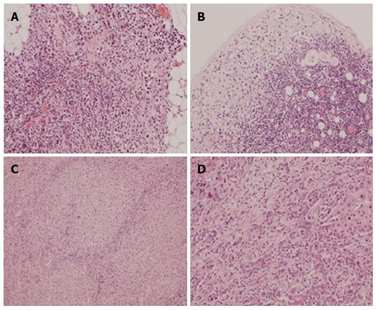
Figure 5 Histopathologic evaluation performed after open D2 gastrectomy combined with liver metastasectomy.
A: Tumor microfocus infiltrations in the peritoneal adipose tissue in the vicinity of the distal surgical margin (obj. 20 ×, HE); B: Metastatic foci in the subcapsular region of the lymph node (obj. 20 ×, HE); C, D: Liver metastasis of gastric carcinoma (obj. 10 ×, 20 ×, HE).
- Citation: Nowacki M, Grzanka D, Zegarski W. Pressurized intraperitoneal aerosol chemotheprapy after misdiagnosed gastric cancer: Case report and review of the literature. World J Gastroenterol 2018; 24(19): 2130-2136
- URL: https://www.wjgnet.com/1007-9327/full/v24/i19/2130.htm
- DOI: https://dx.doi.org/10.3748/wjg.v24.i19.2130


