Copyright
©The Author(s) 2018.
World J Gastroenterol. Mar 14, 2018; 24(10): 1144-1151
Published online Mar 14, 2018. doi: 10.3748/wjg.v24.i10.1144
Published online Mar 14, 2018. doi: 10.3748/wjg.v24.i10.1144
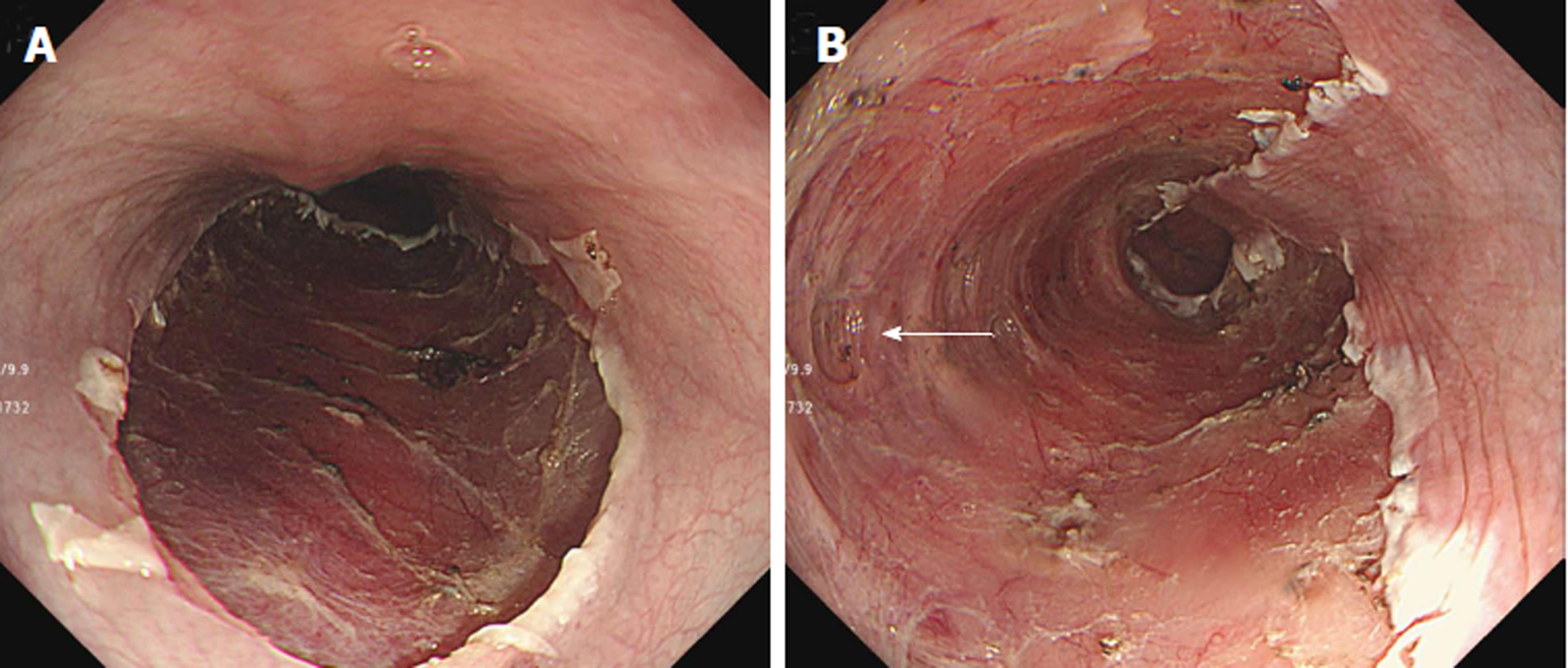
Figure 1 Proper muscle layer exposure during endoscopic submucosal dissection in esophagus.
A: Absent; B: Present.
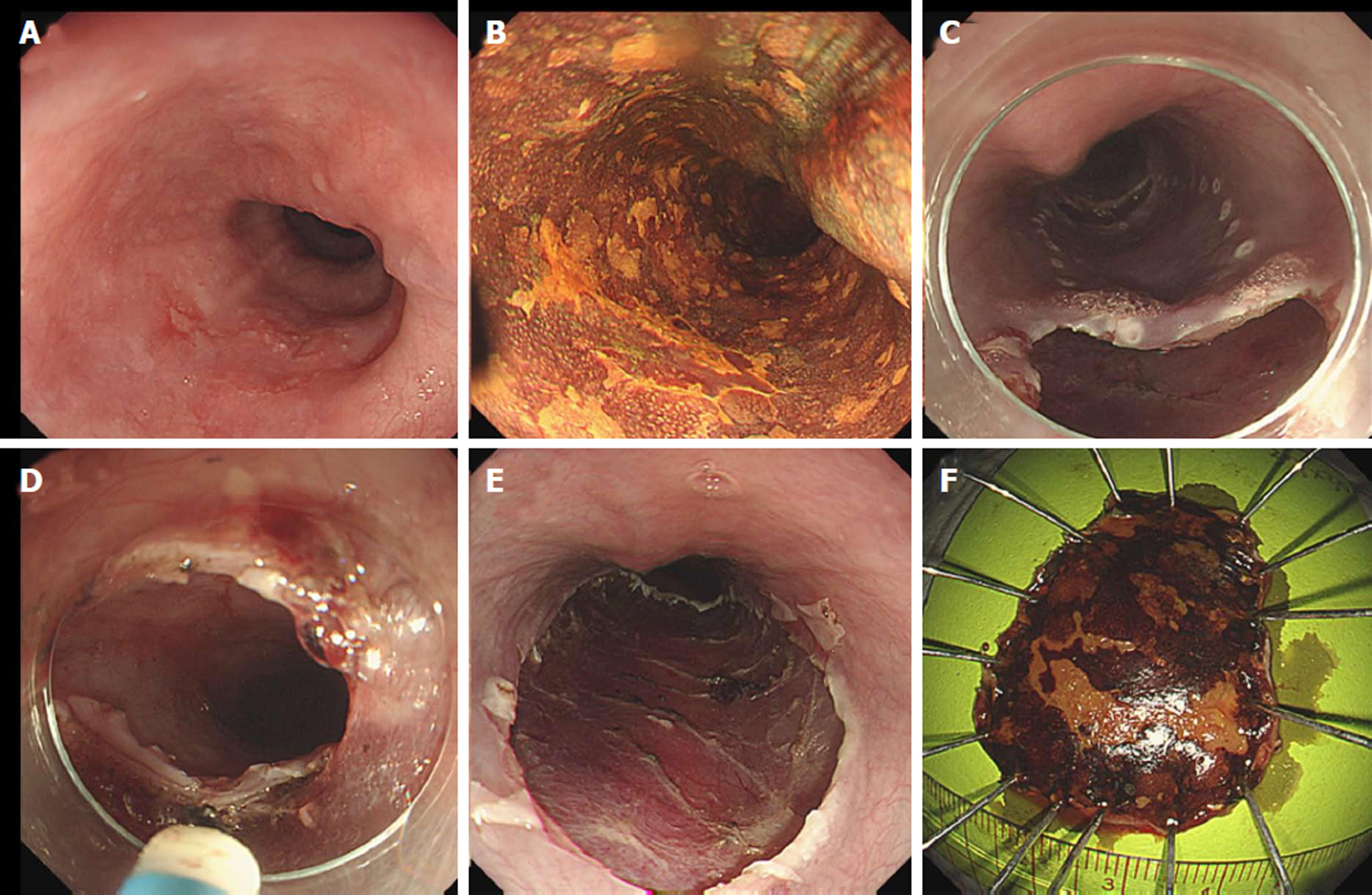
Figure 2 Endoscopic submucosal dissection of a superficial esophageal neoplasm.
A and B: A flat erythematous lesion that is unstained with Lugol’s solution; C and D: Endoscopic submucosal dissection is made with a dual-knife after local submucosal injection; E and F: The lesion is completely resected.
- Citation: Ma DW, Youn YH, Jung DH, Park JJ, Kim JH, Park H. Risk factors of electrocoagulation syndrome after esophageal endoscopic submucosal dissection. World J Gastroenterol 2018; 24(10): 1144-1151
- URL: https://www.wjgnet.com/1007-9327/full/v24/i10/1144.htm
- DOI: https://dx.doi.org/10.3748/wjg.v24.i10.1144


