Copyright
©The Author(s) 2017.
World J Gastroenterol. Dec 21, 2017; 23(47): 8426-8431
Published online Dec 21, 2017. doi: 10.3748/wjg.v23.i47.8426
Published online Dec 21, 2017. doi: 10.3748/wjg.v23.i47.8426
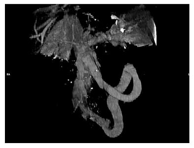
Figure 1 Volume rendering CECT (portal phase).
Showing the giant porto-systemic shunt, patent portal and splenic veins and no gastro-esophageal varices. The shunt extends from the inferior part of the spleno-mesenteric confluence to the left hypogastric vein. Enlarged calibre of the superior mesenteric vein is visible at the confluence.
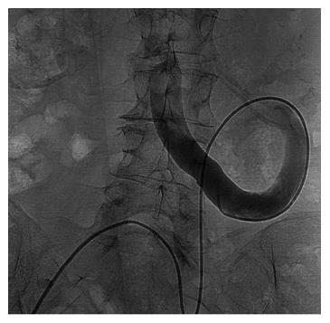
Figure 2 Direct venography from right femoral vein approach, by a 5 Fr diagnostic catheter, of the porto-systemic shunt.
Confirming the presence of the retroperitoneal loop with a hepatofugal flow to the left internal iliac vein.
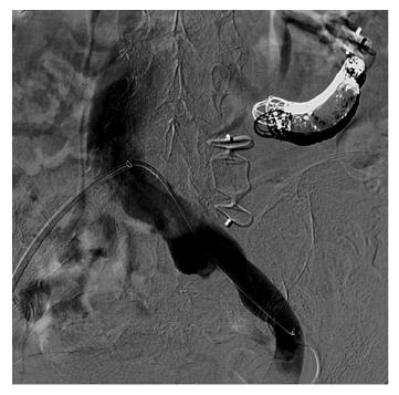
Figure 3 Completion angiography.
Shunt exclusion after endovascular embolization by detachable coils and two plugs and patency of the left internal iliac vein and inferior vena cava.
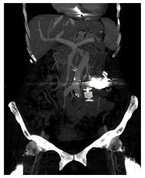
Figure 4 Follow up MIP (maximum intensity projection) computed tomography at 1 mo.
Pointed out the patency of the superior mesenteric, splenic and portal veins. Coils cast and plugs, proximal and distal, completely excluded shunt’s flow. Platinum coils-related artifacts are evident above the nitinol plug. The course of aorta parallel to superior mesenteric vein is depicted.
- Citation: de Martinis L, Groppelli G, Corti R, Moramarco LP, Quaretti P, De Cata P, Rotondi M, Chiovato L. Disabling portosystemic encephalopathy in a non-cirrhotic patient: Successful endovascular treatment of a giant inferior mesenteric-caval shunt via the left internal iliac vein. World J Gastroenterol 2017; 23(47): 8426-8431
- URL: https://www.wjgnet.com/1007-9327/full/v23/i47/8426.htm
- DOI: https://dx.doi.org/10.3748/wjg.v23.i47.8426


