Copyright
©The Author(s) 2017.
World J Gastroenterol. Oct 14, 2017; 23(38): 7047-7053
Published online Oct 14, 2017. doi: 10.3748/wjg.v23.i38.7047
Published online Oct 14, 2017. doi: 10.3748/wjg.v23.i38.7047
Figure 1 Esophagogastroduodenoscopy findings.
A: A Borrmann type II gastric cancer was detected at the antrum; B and C: A 10 mm submucosal tumor-like lesion on the lesser curvature of the upper third of the stomach was detected. Atrophic gastritis O-1 according to Kimura and Takemoto classification was recognized.
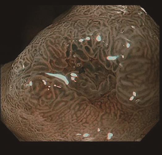
Figure 2 Narrow-band imaging with magnifying endoscopy findings.
There were few irregularities in the microvessel architecture and microsurface structure. Therefore, we diagnosed a regular microvascular pattern and a regular microsurface pattern. The depressed area at the center was expected to be a biopsy scar.
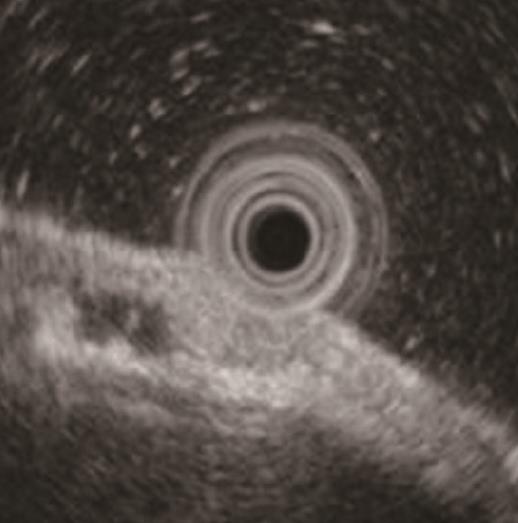
Figure 3 Endoscopic ultrasonography findings.
The tumor slightly invaded the third layer, although it was located mainly in the second layer. Moreover, a hypoechoic mass located in the third layer near the tumor was detected.
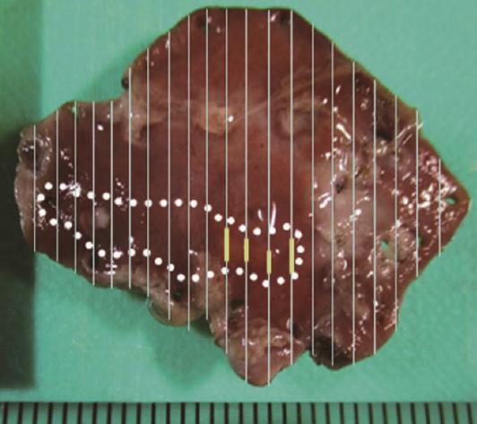
Figure 4 Spread of the gastric adenocarcinoma of fundic gland type and heterotopic gastric glands mapped on the endoscopic submucosal dissection-resected specimen.
Yellow line spread of the gastric adenocarcinoma of fundic gland type (GA-FG), White dotted line spread of heterotopic gastric glands (HGG). The endoscopic submucosal dissection -resected specimen was 30 mm × 20 mm in size. The tumor was slightly elevated and measured 7 mm × 4 mm. The GA-FG and HGG partially overlapped.
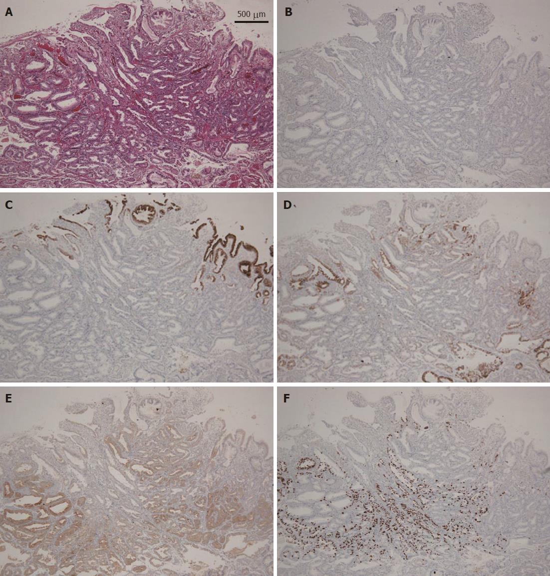
Figure 5 Histopathological and immunohistochemical findings of the endoscopic submucosal dissection-resected specimen.
A: H&E staining. There were atypical cells with mildly enlarged nuclei in the deep layer of the lamina propria mucosae. They mimicked fundic gland cells, mainly chief cells and partially parietal cells. The mucosal surface was covered completely with non-neoplastic foveolar epithelium; thus, the tumor was not exposed to the mucosal surface; B: MUC2 staining: almost negative; C: MUC5AC staining: almost negative except for normal superficial foveolar epithelium; D: MUC6 staining: partially positive and indicating a gastric phenotype; E: Pepsinogen staining: diffusely positive in the deep layer of the lamina propria mucosae, corresponding to gastric adenocarcinoma of fundic gland type, and indicating a differentiation toward chief cells; F: H/K-ATPase staining: scattered positive in the deep layer of the lamina propria mucosae, indicating a differentiation toward parietal cells focally.
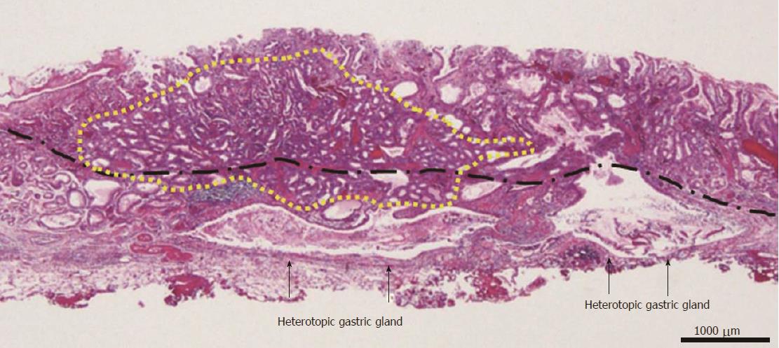
Figure 6 Histopathological findings of the endoscopic submucosal dissection-resected specimen.
Yellow line the gastric adenocarcinoma of fundic gland type (GA-FG), Black line muscularis mucosae. The GA-FG partially invaded the submucosa layer up to 450 μm, although it was located mainly in the deep layer of the lamina propria mucosae. Enlarged ducts of the heterotopic gastric glands were concomitantly observed just under the submucosal lesion of the GA-FG. In addition, the submucosal lesion had a poor stromal reaction.
- Citation: Manabe S, Mukaisho KI, Yasuoka T, Usui F, Matsuyama T, Hirata I, Boku Y, Takahashi S. Gastric adenocarcinoma of fundic gland type spreading to heterotopic gastric glands. World J Gastroenterol 2017; 23(38): 7047-7053
- URL: https://www.wjgnet.com/1007-9327/full/v23/i38/7047.htm
- DOI: https://dx.doi.org/10.3748/wjg.v23.i38.7047


