Copyright
©The Author(s) 2017.
World J Gastroenterol. Aug 21, 2017; 23(31): 5817-5822
Published online Aug 21, 2017. doi: 10.3748/wjg.v23.i31.5817
Published online Aug 21, 2017. doi: 10.3748/wjg.v23.i31.5817
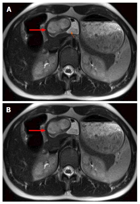
Figure 1 Magnetic resonance imaging findings for case 1, using transverse T2-and T1-weighted magnetic resonance imaging.
A: Transverse T2-weighted magnetic resonance imaging (MRI) showing well circumscribed, endophytic mass (thick red arrow) of the gastric antrum which is partially filled with fluid (thin orange arrow); B: T1-weighted MRI after contrast media application showing homogeneous wall enhancement of the well circumscribed mass (thick red arrow).
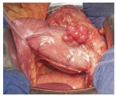
Figure 2 Gross appearance of the plexiform fibromyxoma during surgery (case 2).
The tumor is lobulated, protrudes from the gastric serosa and is located in the lower part (antrum) of the stomach near the pylorus. Underneath the stomach, normal appearing pancreas can be appreciated.
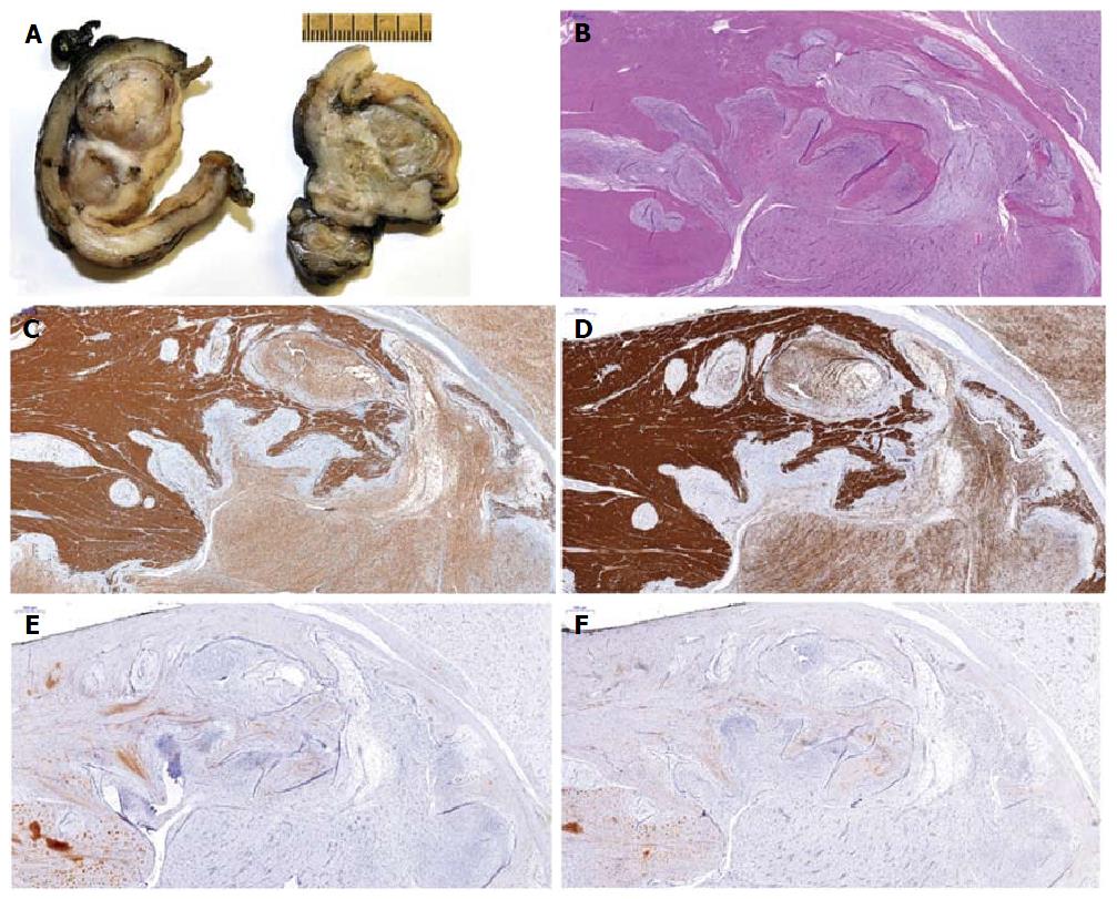
Figure 3 Macroscopic, microscopic and immunohistochemical features of plexiform fibromyxoma.
A: Cut section showing multinodular, solid glistening translucid tumor within the gastric wall and subserosa; B: Histological analysis of the tumor showing composition of ovoid to spindled-shaped cells in a myxoid or fibromyxoid stroma with a plexiform intramural growth pattern; C-F: Immunohistochemical analysis showing positivity for alpha-smooth muscle actin (C) and h-caldesmon (D), but negativity for KIT (E) and DOG1 (F).
- Citation: Szurian K, Till H, Amerstorfer E, Hinteregger N, Mischinger HJ, Liegl-Atzwanger B, Brcic I. Rarity among benign gastric tumors: Plexiform fibromyxoma - Report of two cases. World J Gastroenterol 2017; 23(31): 5817-5822
- URL: https://www.wjgnet.com/1007-9327/full/v23/i31/5817.htm
- DOI: https://dx.doi.org/10.3748/wjg.v23.i31.5817


