Copyright
©The Author(s) 2017.
World J Gastroenterol. Aug 21, 2017; 23(31): 5798-5808
Published online Aug 21, 2017. doi: 10.3748/wjg.v23.i31.5798
Published online Aug 21, 2017. doi: 10.3748/wjg.v23.i31.5798
Figure 1 Preparation of special instruments.
A: The retractors, which are modified from thyroid retractors, could be adapted to the curvature of the pelvis manually. Red dots show the turning point of the retractor during transanal operation; B: An anal dilator with a papilionaceous fixture from a stapler device for hemorrhoids, which was placed after completion of intersphincteric resection.
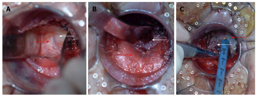
Figure 2 Dissection of the distal mesorectum in retrograde transanal total mesorectal excision.
A: Bilateral mobilization of the distal rectum. Dissection is started along the natural boundary between the surface of the levator ani muscle and mesorectum toward the pelvic cavity assisted. The two retractors are inserted into this space and used to expand it. The distance of bilateral mobilization toward the pelvic cavity could reach 10 cm according to the length of the retractors; B: Posterior mobilization of the distal rectum. The hiatal ligament is cut off after sharp dissection along the natural boundary between the surface of the levator ani muscle and mesorectum with an electrocautery or ultracision harmonic scalpel; C: Anterior mobilization of the distal rectum. The rectourethral muscle is cut off, and the Denonvilliers fascia is sharply dissected between the anterior and posterior lobes. White arrows indicate the rectum and its mesorectum, and red arrow indicates the levator ani muscle or Denonvilliers fascia.
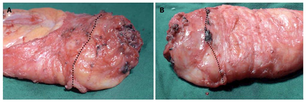
Figure 3 Specimen was examined by a pathologist.
A: The anterior side of specimen; B: The posterior side of specimen. The black dotted line shows the boundary of transabdominal total mesorectal excision (TME) and retrograde transanal TME. The lower rectum had a smoother mesorectum surface compared with the upper rectum. TME: Total mesorectal excision.
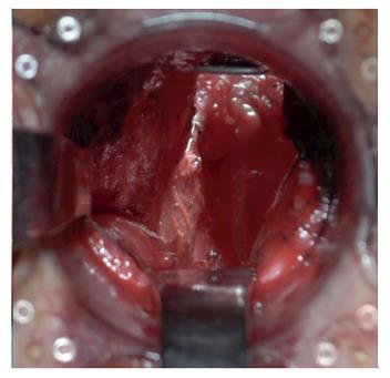
Figure 4 Completion status of the surgery on the pelvic floor (the location of the distal rectum) can be clearly observed transanally after removing the specimen.
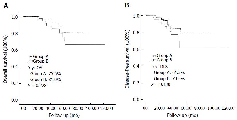
Figure 5 Survival curve for 115 male patients with low rectal cancer.
A: Overall survival of the two groups; B: Disease-free survival of the two groups.
- Citation: Xu C, Song HY, Han SL, Ni SC, Zhang HX, Xing CG. Simple instruments facilitating achievement of transanal total mesorectal excision in male patients. World J Gastroenterol 2017; 23(31): 5798-5808
- URL: https://www.wjgnet.com/1007-9327/full/v23/i31/5798.htm
- DOI: https://dx.doi.org/10.3748/wjg.v23.i31.5798


