Copyright
©The Author(s) 2017.
World J Gastroenterol. Aug 21, 2017; 23(31): 5700-5712
Published online Aug 21, 2017. doi: 10.3748/wjg.v23.i31.5700
Published online Aug 21, 2017. doi: 10.3748/wjg.v23.i31.5700
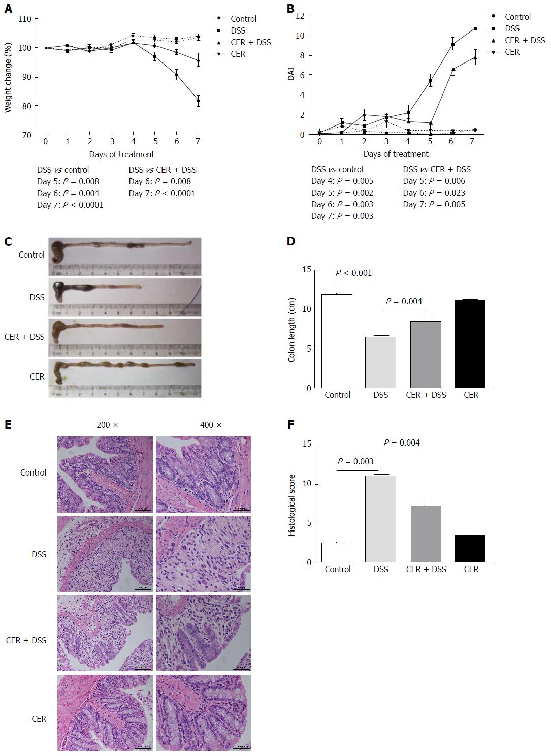
Figure 1 Treatment with S.
japonicum cercariae results in reduced susceptibility to dextran sodium sulfate-induced colitis in mice. Mice were infected with 20 ± 2 S. japonicum cercariae percutaneously, and experimental colitis was induced by administration of 3% DSS at 4 wk post infection. A: Weight change in percent; B: DAI based on weight change, stool characteristics and bleeding; C and D: Colon length; E: H and E staining was performed in colonic sections (original magnification, × 200 and × 400); F: HS was scored as described in Materials and Methods. n = 6. The mice infected with S. japonicum cercariae presented longer colons, decreased weight loss and lower DAI and HS after being induced with DSS compared to those without parasite infection. DSS: Dextran sodium sulfate treatment alone; CER + DSS: Infection with S. japonicum before DSS treatment; CER: S. japonicum infection alone.
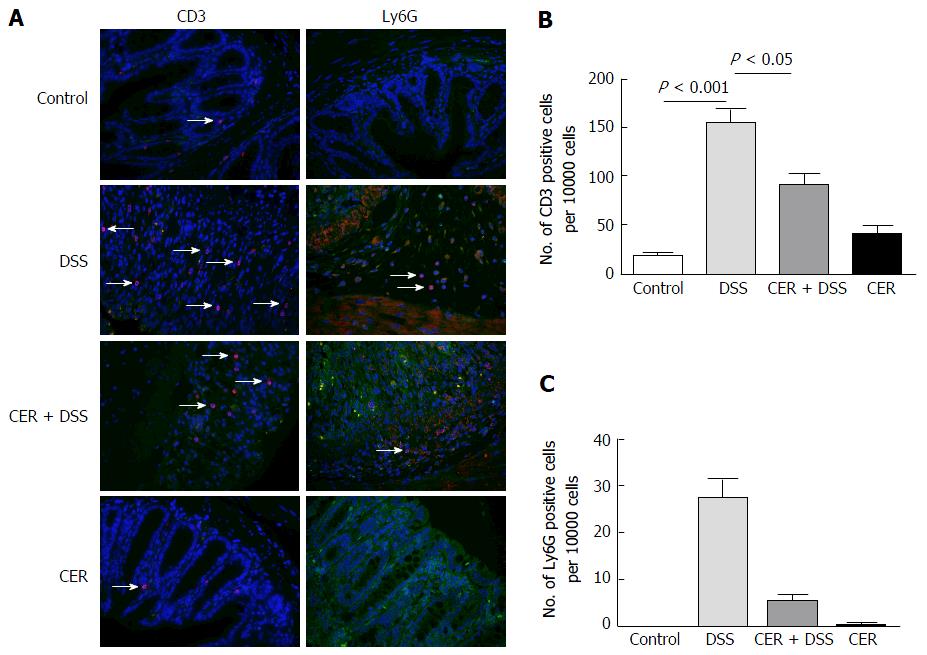
Figure 2 Infection with S.
japonicum decreases infiltration of inflammatory cells in colon tissues. A: Immunofluorescence staining of CD3 and Ly6G in the colon sections; B and C: Respective quantification of CD3 and Ly6G positive cells. n = 3. DSS: Dextran sodium sulfate treatment alone; CER + DSS: Infection with S. japonicum before DSS treatment; CER: S. japonicum infection alone.
Figure 3 Administration of S.
japonicum leads to lowered inflammatory cytokines in dextran sodium sulfate-induced colitis mice. A: The production of IL-2, IL-6, IL-10, IL-17A, TNFα and IFN-γ in blood samples was detected by CBA and flow cytometry; B: IL-1β, IL-6, IL-10, IL-17a, IFN-γ, TNFα and TGFβ levels in colon tissues were tested by RT-PCR. n = 3 or 4. Exposure to S. japonicum protected mice from DSS-induced colitis with down-regulation of the Th1/Th2/Th17 pathway. DSS: Dextran sodium sulfate treatment alone; CER + DSS: Infection with S. japonicum before DSS treatment; CER: S. japonicum infection alone.
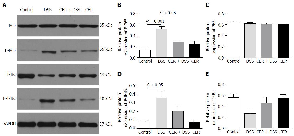
Figure 4 Infection with S.
japonicum decreases the expression of phosphorylated-p65 in mice treated with dextran sodium sulfate. A: The expression of P-P65 and P65 in colon tissues was examined by Western blot; B and C: Respective quantification of P-P65 and P65; D and E: Respective quantification of P-IKBα and IKBα. n = 3. DSS: Dextran sodium sulfate treatment alone; CER + DSS: Infection with S. japonicum before DSS treatment; CER: S. japonicum infection alone.
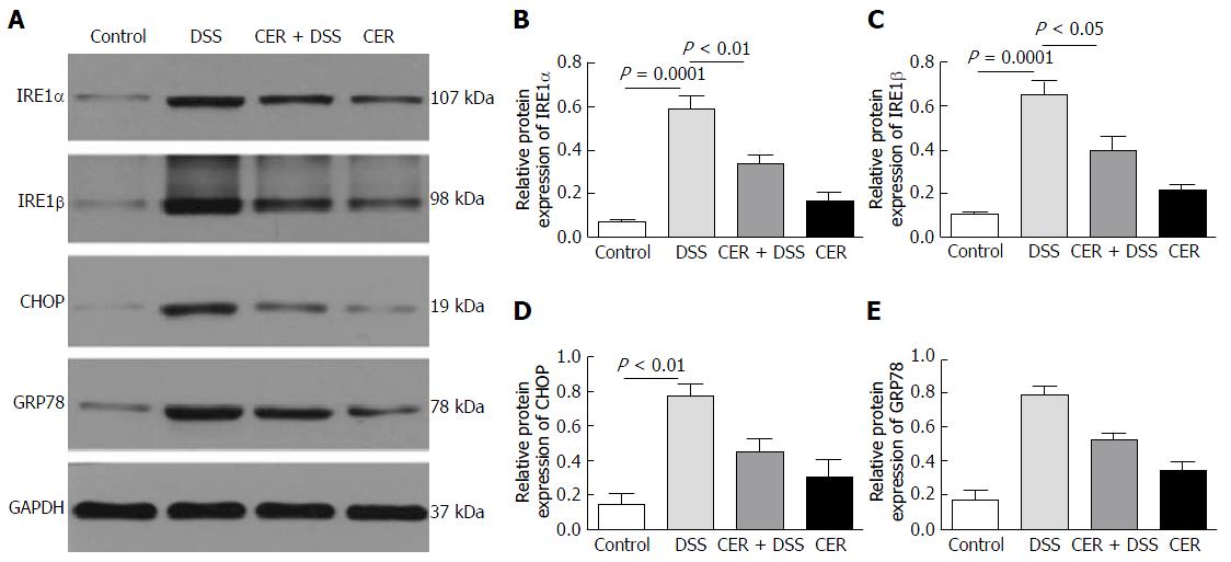
Figure 5 Endoplasmic reticulum stress markers are downregulated in mice induced with dextran sodium sulfate after infection.
A: The expression of IRE1α, IRE1β, GRP78 and CHOP in colon tissues was examined by Western blot; B-E: Respective quantification of IRE1α, IRE1β, GRP78 and CHOP. n = 4. DSS: Dextran sodium sulfate treatment alone; CER + DSS: Infection with S. japonicum before DSS treatment; CER: S. japonicum infection alone.
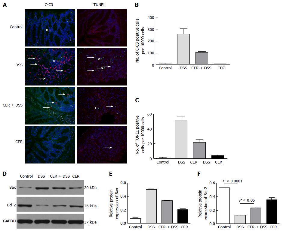
Figure 6 Apoptosis in colitic mice after infection with S.
japonicum. A: Immunofluorescence staining of cleaved-caspase3 (C-C3) and TUNEL staining; B and C: Respective quantification of C-C3 and TUNEL positive cells; D: Bax and Bal-2 were tested by Western blot; E and F: Respective quantification of Bax and Bal-2. n = 3. DSS: Dextran sodium sulfate treatment alone; CER + DSS: Infection with S. japonicum before DSS treatment; CER: S. japonicum infection alone.
- Citation: Liu Y, Ye Q, Liu YL, Kang J, Chen Y, Dong WG. Schistosoma japonicum attenuates dextran sodium sulfate-induced colitis in mice via reduction of endoplasmic reticulum stress. World J Gastroenterol 2017; 23(31): 5700-5712
- URL: https://www.wjgnet.com/1007-9327/full/v23/i31/5700.htm
- DOI: https://dx.doi.org/10.3748/wjg.v23.i31.5700


