Copyright
©The Author(s) 2017.
World J Gastroenterol. Jun 21, 2017; 23(23): 4243-4251
Published online Jun 21, 2017. doi: 10.3748/wjg.v23.i23.4243
Published online Jun 21, 2017. doi: 10.3748/wjg.v23.i23.4243
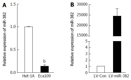
Figure 1 miR-382 was down-regulated in the esophageal squamous cell carcinoma cell line Eca109.
A: The expression of miR-382 in Het-1A cells and Eca109 cells was examined by RT-qPCR; B: The relative expression level of miR-382 after introduction of either lentivirus-mediated miR-382 (LV-miR-382) or lentivirus-mediated scrambled control (LV-Con) into Eca109 cells was examined by RT-qPCR. The data in A and B is expressed as mean ± SD of three independent experiments. bP < 0.01, vs control. ESCC: Esophageal squamous cell carcinoma.
Figure 2 miR-382 inhibited Eca109 cell proliferation and colony formation.
A: The proliferation of miR-382 overexpressed Eca109 cells and LV-Con control cells was measured by MTT. The Y-axis represents the relative number of viable cells presented as mean ± SD, bP < 0.01; B: Representative photomicrographs show the colony formation in cultured LV-Con Eca109 cells and LV-miR-382 Eca109; C: Data is presented as mean ± SD, bP < 0.01.
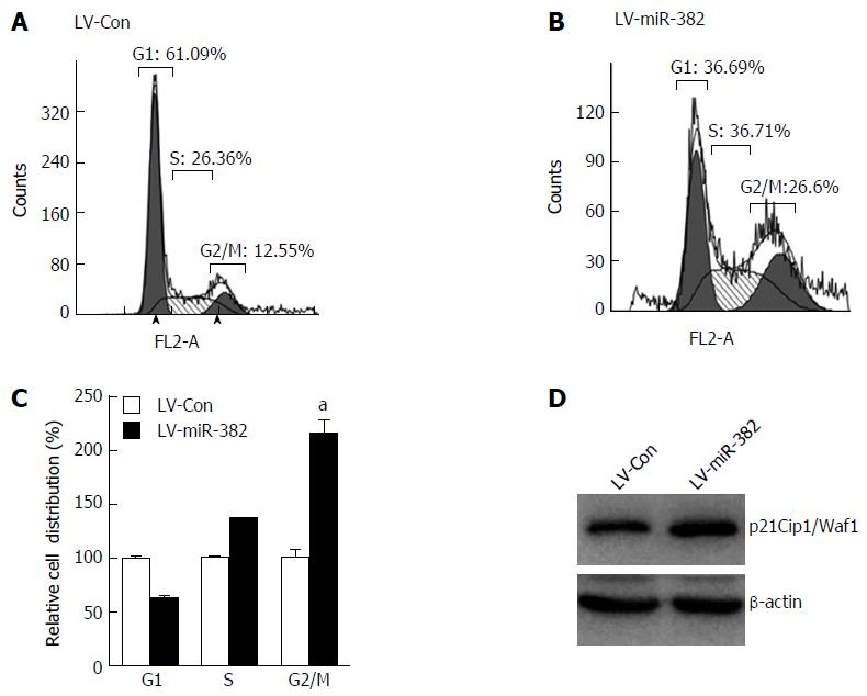
Figure 3 miR-382 induced cell cycle arrest at G2/M phase in Eca109 cells.
A and B: Cell cycle distribution of miR-382 overexpressed Eca109 cells and control cells was analyzed by flow cytometry at post-culture 48 h; C: Relative cell distribution percentage of LV-miR-382 expressed Eca109 cells to LV-Con control cells in each cell cycle phase. The data presented is mean ± SD of three independent experiments. aP < 0.05, vs control; D: Protein expression level of p21Cip1/Waf1 was examined in LV-Con Eca109 cells and LV-miR-382 Eca109 cells. β-actin was as a loading control.
Figure 4 miR-382 promoted the apoptotic process in Eca109 cells.
A: Apoptosis of LV-Con Eca109 cells and LV-miR-382 Eca109 cells was a negative cell cycle regulator, as measured using flow cytometry; B: Apoptotic cell values were expressed as mean ± SD of three experiments. aP < 0.05 and bP < 0.01, vs control.
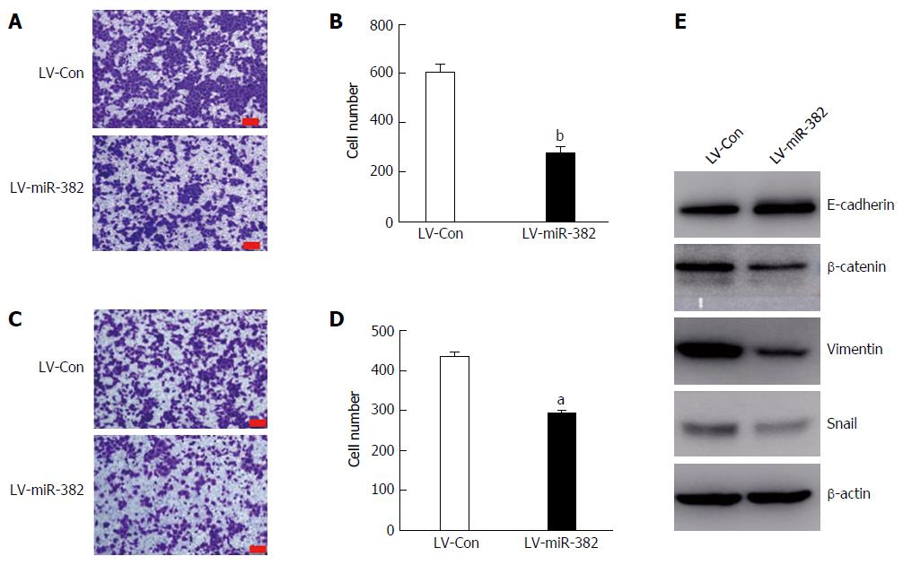
Figure 5 Overexpression miR-382 inhibited migration, invasion and epithelial-mesenchymal transition in Eca109 cells.
A: Representative photomicrographs showed migrated cells stained with crystal violet after post-culture for 24 h in the transwell migration assay. The bars represent 200 μm; B: The migrated cells in five different fields were counted and the Y-axis represents the migrated cell number from three independent experiments. Data is presented as mean ± SD, bP < 0.01; C: Representative photomicrographs showing invaded cells stained with crystal violet after post-culture for 24 h in the transwell invasion assay. The bars are 200 μm; D: The invaded cells in five different fields were counted and the Y-axis represents the invaded cell number from three independent experiments. Data are presented as mean ± SD, aP < 0.05; E: Western blot analysis of the expression of epithelial marker E-cadherin, and mesenchymal markers β-catenin, vimentin and snail in Eca109 cells infected with LV-miR-382 or LV-Con(c). β-actin served as a loading control.
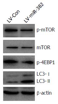
Figure 6 miR-382 inhibited mTOR and 4E-BP1 phosphorylation and activated autophagy.
p-mTOR, mTOR, p-4E-BP1 and LC3 were evaluated by western blot. Experiments were performed in triplicate. Equal protein loading was confirmed by β-actin.
- Citation: Feng J, Qi B, Guo L, Chen LY, Wei XF, Liu YZ, Zhao BS. miR-382 functions as a tumor suppressor against esophageal squamous cell carcinoma. World J Gastroenterol 2017; 23(23): 4243-4251
- URL: https://www.wjgnet.com/1007-9327/full/v23/i23/4243.htm
- DOI: https://dx.doi.org/10.3748/wjg.v23.i23.4243


