Copyright
©The Author(s) 2017.
World J Gastroenterol. Jun 21, 2017; 23(23): 4140-4145
Published online Jun 21, 2017. doi: 10.3748/wjg.v23.i23.4140
Published online Jun 21, 2017. doi: 10.3748/wjg.v23.i23.4140
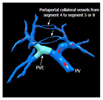
Figure 1 Bilateral cross-portal circulation after portal vein embolization.
Red arrows indicate blood flow to the S8 or S5 from the collateral vessels. PV: Portal vein; PVE: Portal vein embolization.
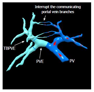
Figure 2 Terminal branches portal vein embolization of S8 and S5.
The communicating portal vein branches were interrupted without liver parenchyma division (red arrows indicate that blood flow to the S8 or S5 was interrupted, and blue dotted lines indicate the boundary between PVE and TBPVE). PV: Portal vein; PVE: Portal vein embolization; TBPVE: Terminal branches portal vein embolization.
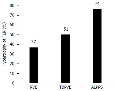
Figure 3 Mean future liver remnant hypertrophy with portal vein embolization, terminal branches portal vein embolization, and associating liver partition and portal vein ligation for staged hepatectomy.
FLR: Future liver remnant; PVE: Portal vein embolization; TBPVE: Terminal branches portal vein embolization; ALPPS: Associating liver partition and portal vein ligation for staged hepatectomy.
- Citation: Peng SY, Wang XA, Huang CY, Zhang YY, Li JT, Hong DF, Cai XJ. Evolution of associating liver partition and portal vein ligation for staged hepatectomy: Simpler, safer and equally effective methods. World J Gastroenterol 2017; 23(23): 4140-4145
- URL: https://www.wjgnet.com/1007-9327/full/v23/i23/4140.htm
- DOI: https://dx.doi.org/10.3748/wjg.v23.i23.4140


