Copyright
©The Author(s) 2017.
World J Gastroenterol. Jun 7, 2017; 23(21): 3928-3933
Published online Jun 7, 2017. doi: 10.3748/wjg.v23.i21.3928
Published online Jun 7, 2017. doi: 10.3748/wjg.v23.i21.3928
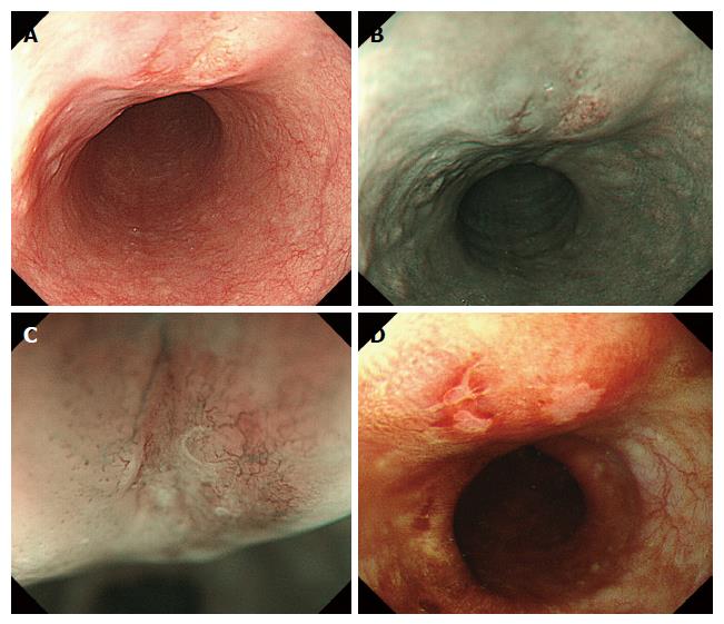
Figure 1 Endoscopic appearance.
A: White-light conventional endoscopy showed a submucosal tumor-like lesion. There were two reddish depressed areas on the surface of the tumor; B: The lesion appeared as brownish areas on NBI; C: Magnification endoscopy with NBI revealed irregular loop-shaped microvessels coexisting with irregularly branched thick non-looped vessels in depressed areas, where Lugol’s iodine (D) showed negative staining.
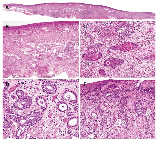
Figure 2 Perspective view of the resected specimen.
A: Variably sized glands are diffusely dispersed accompanying stromal fibrosis in the mucosal and submucosal layers. In the deepest zone, some solid nests without a luminal structure are evident; B: Invasive carcinoma is composed of round or oval-shaped glands and irregularly dilated glands; C: Invasive tumor nests of keratinizing squamous cell carcinoma are observed in the deepest part of the lesion; D: A higher-power view shows that the glands are have two cell layers. Most cells do not exhibit enough atypia to support a diagnosis of dysplasia; E: The underlying carcinoma component is focally continuous with the surface covering epithelial layer. Although the covering epithelium is obviously thinned compared with surrounding epithelium, we found no significant atypia in the area continuous with the invasive carcinoma. B and E: Magnification 100 ×; C and D Magnification 200 ×.
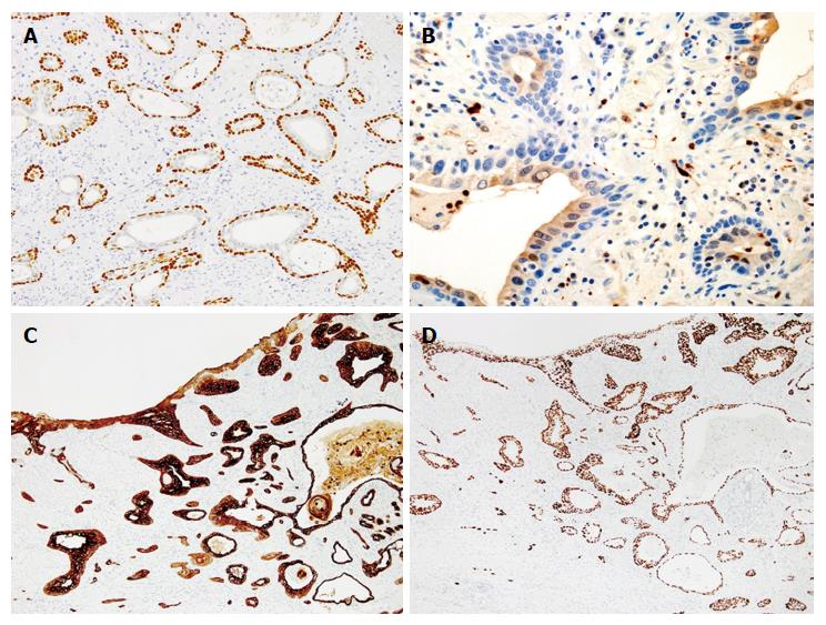
Figure 3 Immunohistochemical examinations.
A: p63 (magnification 200 ×); B: S-100 (magnification 400 ×); C: CK7 (magnification 100 ×); D: p53 (magnification 100 ×). The outer layer cells of the neoplastic tubules were reactive for p63 and CK7, but negative for S100. The inner layer cells were immunopositive for S100, CK7, but negative for p63. Strong expressions of p53 and CK7 were observed in the small area of the surface epithelium connecting with the invasive carcinoma component. Both the inner and outer layer cells in the invasive component also overexpressed p53 protein.
- Citation: Tamura H, Saiki H, Amano T, Yamamoto M, Hayashi S, Ando H, Doi R, Nishida T, Yamamoto K, Adachi S. Esophageal carcinoma originating in the surface epithelium with immunohistochemically proven esophageal gland duct differentiation: A case report. World J Gastroenterol 2017; 23(21): 3928-3933
- URL: https://www.wjgnet.com/1007-9327/full/v23/i21/3928.htm
- DOI: https://dx.doi.org/10.3748/wjg.v23.i21.3928


