Copyright
©The Author(s) 2017.
World J Gastroenterol. May 28, 2017; 23(20): 3752-3757
Published online May 28, 2017. doi: 10.3748/wjg.v23.i20.3752
Published online May 28, 2017. doi: 10.3748/wjg.v23.i20.3752
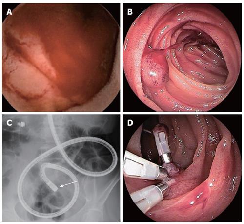
Figure 1 Evaluation of endoscopic findings (case 1).
Video capsule endoscopy (A) and double-enteroscopy (B and C) show a raised lesion with smooth surface in the upper jejunum, and double-balloon enterscopy showed spout bleeding of the lesion. The lesion in the jejunum was disclosed 29 min after capsule ingestion (pylonus passage at 16 min) (A). Detailed localization of the target lesion using fluoroscopy is shown by the end of endoscopic insertion (arrow) (C). The lesion underwent endoscopic hemostatic clipping (D).
Figure 2 Surgical and pathological finding (case 1).
The surgical finding on single port laparoscopic surrey shows an objective site with India link tattooing (A). Surgical specimen form the small intestine. In cluding the lesion indicated with India link shows a whole view of the resected lesion (B). Histological (h-e stain) in the resected specimen show different-sized blood vessels circumferentially proliferated from the mucosa to submucosa. Inset show different-sized distended blood vessels circumferentially proliferated from the mucosa to submucosa. Inset shows a low-power filed view (C).
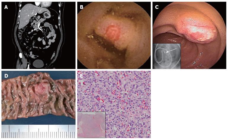
Figure 3 Evaluation of clinical finding (case 2).
Early-phase contrast-enhanced computed tomography reveals small nodule enhancement in the ileum (arrow) (A). Video capsule endoscopy (B) and double-balloon enteroscopy (C) show a submucosal tumor-like raised lesion with central erosion in the lower ileum. The lesion in the jejunum was disclosed 145 min after capsule ingestion (pylorus passage at 140 min) (B), inset indicates fluoroscopic localization of target at the end of the endoscope insertion (arrow) (C). Surgical specimen from the small intestine, including the indicated lesion with India ink tattooing (D). Histological finding (H-E stain) in the resected specimen show cirumferential capillary growth without atypia from the mucosa to the muscle. Inset shows a low-power filed view (E).
- Citation: Takase N, Fukui K, Tani T, Nishimura T, Tanaka T, Harada N, Ueno K, Takamatsu M, Nishizawa A, Okamura A, Kaneda K. Preoperative detection and localization of small bowel hemangioma: Two case reports. World J Gastroenterol 2017; 23(20): 3752-3757
- URL: https://www.wjgnet.com/1007-9327/full/v23/i20/3752.htm
- DOI: https://dx.doi.org/10.3748/wjg.v23.i20.3752


