Copyright
©The Author(s) 2017.
World J Gastroenterol. May 28, 2017; 23(20): 3675-3683
Published online May 28, 2017. doi: 10.3748/wjg.v23.i20.3675
Published online May 28, 2017. doi: 10.3748/wjg.v23.i20.3675
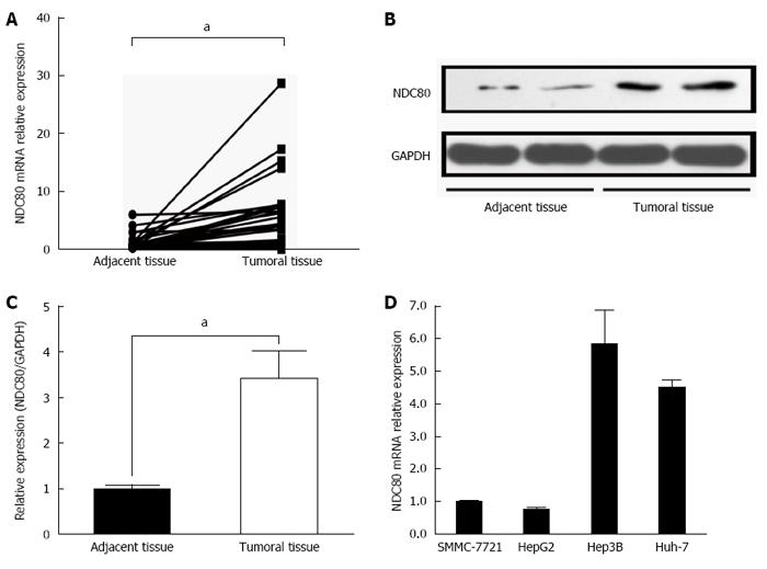
Figure 1 Reverse transcription polymerase chain reaction results for NDC80 mRNA expression.
A: Expression levels of NDC80 mRNA in HCC (n = 47) and paired adjacent tissue samples (n = 47); B: NDC80 protein expression in HCC and paired adjacent tissue samples was determined by western blotting; C: Gray value analysis of western blot experiments, and data was normalized against GAPDH; D: NDC80 mRNA expression varied among SMMC-7721, HepG2, Hep3B and Hun-7 cell lines. GAPDH was used as an internal control. Statistical significance was assessed by paired t tests. Error bar indicates SD (aP < 0.001 vs control).
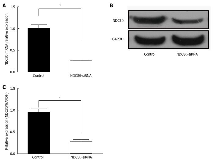
Figure 2 Interference efficiency 72 h after transfection.
A: After lentiviral transfection, relative NDC80 mRNA expression was significantly inhibited in the SMMC-7721 NDC80-siRNA silenced cells as compared to SMMC-7721 negative control cells by RT-PCR; B: Western blotting of NDC80-depletion efficiency in SMMC-7721 cells; C: Gray value analysis of western blotting, and data were normalized against GAPDH. GAPDH was used as an internal control. Statistical significance was assessed by two-tailed Student’s t test. Error bar indicates SD (cP < 0.01 vs control; aP < 0.001 vs control).
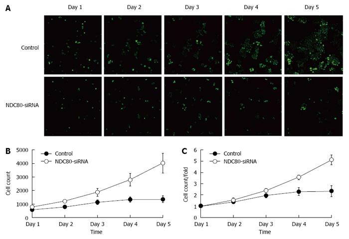
Figure 3 Cell proliferation analysis by green fluorescent protein-based imaging and MTT assay.
A: After lentiviral transfection of SMMC-7721 cells, cell proliferation was significantly inhibited in NDC80-siRNA-silenced cells as compared to the control cells according to green fluorescent protein-based Cellomics ArrayScan VTI imaging; B: After lentiviral transfection of SMMC-7721 cells, MTT assays were performed at the days indicated to show the proliferation of SMMC-7721 cells. The MTT value ratio was significantly reduced in the NDC80-siRNA-silenced cells as compared to the control cells.
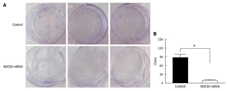
Figure 4 Effects of the silencing of NDC80 on SMMC-7721 cell colony formation.
A: After lentiviral transfection of SMMC-7721 cells, the NDC80-siRNA-silenced cells displayed a significantly reduced number of cell colonies compared to control cells. Colonies were stained with crystal violet. The whole plate fields were photographed and presented. The number of cell colonies of triplicate values in a representative experiment was counted; B: Statistical significance was assessed by two-tailed Student’s t test. Error bar indicates SD (aP < 0.001 vs control).
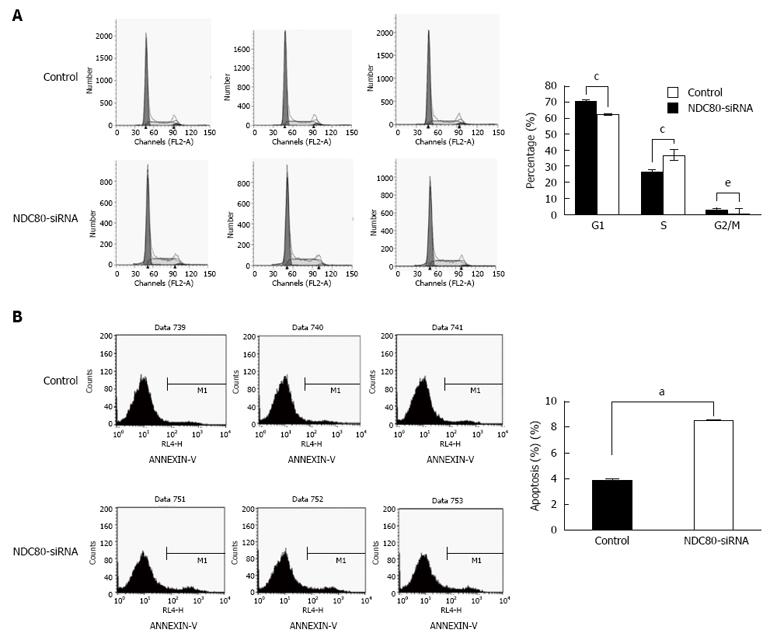
Figure 5 Effects of the silencing of NDC80 on SMMC-7721 cell cycle distribution and apoptosis.
A: After lentiviral transfection of SMMC-7721 cells, cell cycle assessment showed that knockdown of NDC80 in SMMC-7721 cells induced accumulation in S-phase. The percentages of cells in different phases are shown as the mean ± SD of three independent experiments; B: Apoptotic rates were analyzed by Annexin V-FITC/PI assay. Apoptosis was significantly increased in NDC80-siRNA-silenced cells as compared to control cells. Statistical significance was assessed by two-tailed Student’s t test. Error bar indicates SD (eP < 0.05; cP < 0.01; aP < 0.0001, vs control).
- Citation: Ju LL, Chen L, Li JH, Wang YF, Lu RJ, Bian ZL, Shao JG. Effect of NDC80 in human hepatocellular carcinoma. World J Gastroenterol 2017; 23(20): 3675-3683
- URL: https://www.wjgnet.com/1007-9327/full/v23/i20/3675.htm
- DOI: https://dx.doi.org/10.3748/wjg.v23.i20.3675


