Copyright
©The Author(s) 2017.
World J Gastroenterol. May 28, 2017; 23(20): 3655-3663
Published online May 28, 2017. doi: 10.3748/wjg.v23.i20.3655
Published online May 28, 2017. doi: 10.3748/wjg.v23.i20.3655
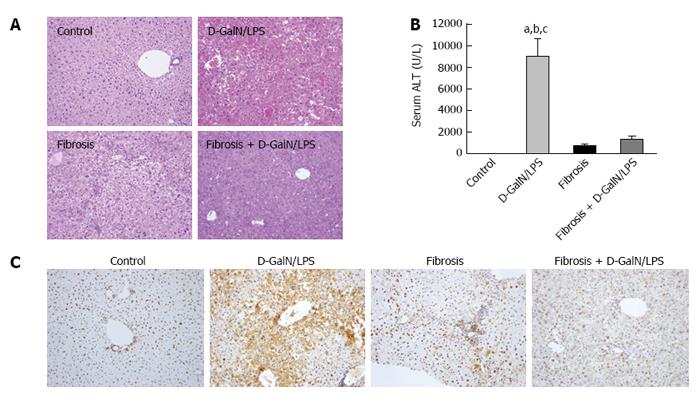
Figure 1 Inhibition of High mobility group box 1 expression is closely associated with the injury resistance in the setting of liver fibrosis.
Control and fibrotic mice (treated with CCl4 for 6 wk) were challenged with a lethal dose of D-GalN (1 mg/g)/LPS (50 ng/g), and hepatic damage was assessed by histology (A: HE staining; original magnification, × 200) and serum ALT levels (B). aP < 0.05 vs the control group, bP < 0.05 vs the fibrosis group, cP < 0.05 vs the fibrosis + D-GalN/LPS group. The expression of HMGB1 was determined by immunohistochemical staining (C: original magnification, × 200). Data are expressed as mean ± SEM. CCl4: Carbon tetrachloride; D-GalN: D-galactosamine; HMGB1: High mobility group box 1; LPS: Lipopolysaccharide.
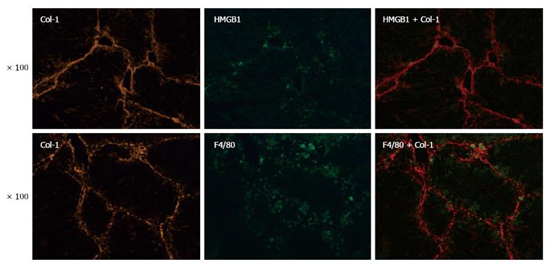
Figure 2 Kupffer cells may be involved in High mobility group box 1-mediated injury resistance.
The expression and localization of F4/80 (a surrogate marker of KCs), HMGB1, and Col-1 were determined by immunofluorescence staining (original magnification, × 100). Col-1: Type I collagen; HMGB1: High mobility group box 1; KCs: Kupffer cells.
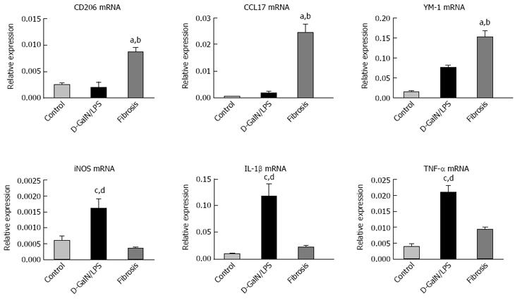
Figure 3 Kupffer cells in the fibrotic liver exhibit predominantly a M2-like activation.
KCs were isolated from the livers of normal, acutely injured (D-GalN/LPS) and fibrotic mice, and M1 and M2 gene signatures were then determined by quantitative real-time PCR. Data are expressed as mean ± SEM. aP < 0.05 vs the control group, bP < 0.05 vs the D-GalN/LPS group, cP < 0.05 vs the control group, dP < 0.05 vs the fibrosis group. D-GalN: D-galactosamine; KCs: Kupffer cells; LPS: Lipopolysaccharide.
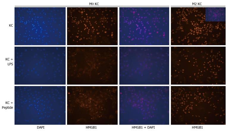
Figure 4 Translocation of High mobility group box 1 triggered by lipopolysaccharide or High mobility group box 1 peptide is inhibited in M2-like Kupffer cells.
KCs were isolated from the livers of control and fibrotic mice, and then treated with LPS (10 ng/mL) or HMGB1 peptide (30 μg/mL). The translocation of HMGB1 in KCs was analyzed by immunofluorescence staining (original magnification, × 200). HMGB1: High mobility group box 1; KCs: Kupffer cells; LPS: Lipopolysaccharide.
- Citation: Zheng QF, Bai L, Duan ZP, Han YP, Zheng SJ, Chen Y, Li JS. M2-like Kupffer cells in fibrotic liver may protect against acute insult. World J Gastroenterol 2017; 23(20): 3655-3663
- URL: https://www.wjgnet.com/1007-9327/full/v23/i20/3655.htm
- DOI: https://dx.doi.org/10.3748/wjg.v23.i20.3655


