Copyright
©The Author(s) 2017.
World J Gastroenterol. Mar 14, 2017; 23(10): 1780-1786
Published online Mar 14, 2017. doi: 10.3748/wjg.v23.i10.1780
Published online Mar 14, 2017. doi: 10.3748/wjg.v23.i10.1780
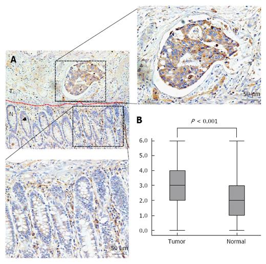
Figure 1 Interleukin-6 expression is elevated in human colorectal cancer tissue samples.
A: Individual FFPE sections demonstrated that colorectal cancer cells had invaded underneath the colorectal mucosa. The IL-6 expression level was elevated in colorectal tumour tissues compared to adjacent non-tumour tissues. “T” refers to tumour tissue, and “N” indicates adjacent non-tumour tissue from the same patient; B: IL-6 expression scores are shown as box plots, with the horizontal lines representing the median, the bottom and top of the boxes representing the 25th and 75th percentiles, respectively, and the vertical bars representing the range of data.
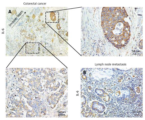
Figure 2 Interleukin-6 expression is associated with invasion depth and lymph node metastasis in colorectal cancer.
A: Increased expression of IL-6 at the invasive front of human CRC samples compared with tumour centre; B: The tumor cells in lymph node metastases were also IL-6-immunopositive. IL: Interleukin.
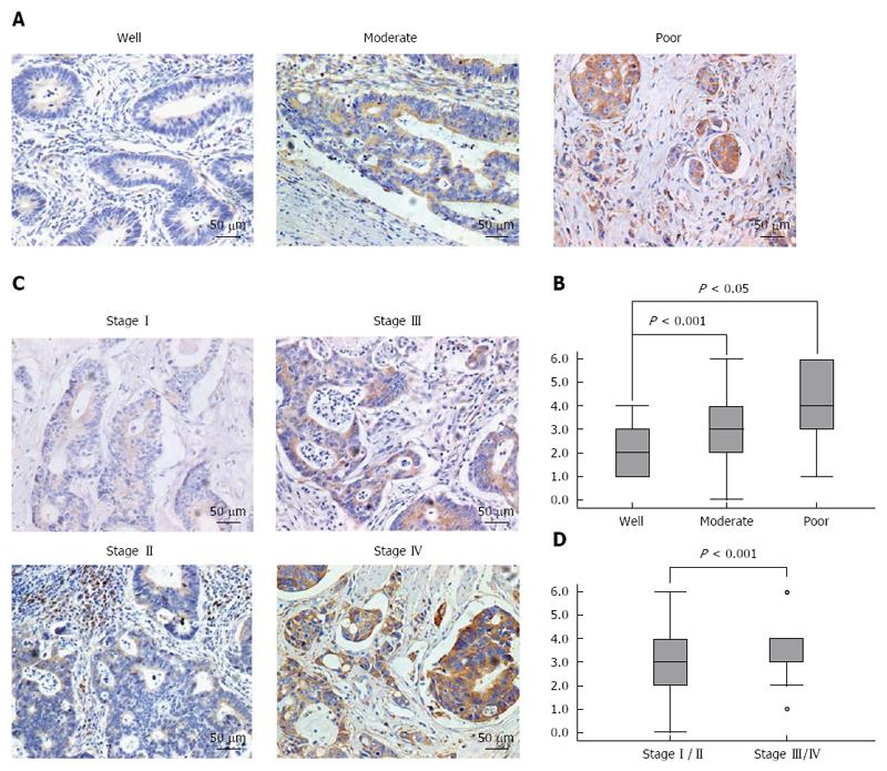
Figure 3 Interleukin-6 expression is significantly correlated with histological differentiation and TNM stage.
A: Poorly differentiated colorectal tumours showed higher expression of IL-6 than moderately and well differentiated colorectal tumours; B: Comparison of immunohistochemical scores of IL-6 according to histological differentiation (P < 0.05); C: Strong immunoreactivity was more likely to present with advanced-stage (stage III/IV) CRC compared to early-stage (stage I/II); D: Comparison of immunohistochemical scores of IL-6 according to TNM stage (P < 0.001).
- Citation: Zeng J, Tang ZH, Liu S, Guo SS. Clinicopathological significance of overexpression of interleukin-6 in colorectal cancer. World J Gastroenterol 2017; 23(10): 1780-1786
- URL: https://www.wjgnet.com/1007-9327/full/v23/i10/1780.htm
- DOI: https://dx.doi.org/10.3748/wjg.v23.i10.1780


