Copyright
©The Author(s) 2016.
World J Gastroenterol. Mar 7, 2016; 22(9): 2855-2860
Published online Mar 7, 2016. doi: 10.3748/wjg.v22.i9.2855
Published online Mar 7, 2016. doi: 10.3748/wjg.v22.i9.2855
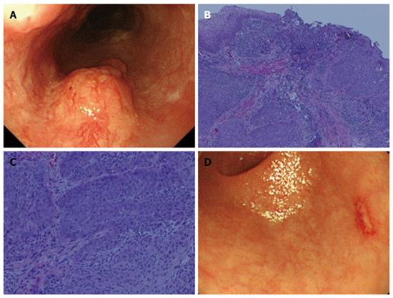
Figure 1 Esophagogastroduodenoscopy findings.
A: An elevated lesion 20 mm in size was detected in the left wall of the middle thoracic esophagus. The endoscopic findings indicated that the lesion was esophageal cancer with a depth of SM2; B, C: Biopsy specimens revealed moderately differentiated squamous cell carcinoma (hematoxylin-eosin stain; magnification: B × 40, C × 200); D: A small erosion with surrounding reddish mucosa was also found in the posterior wall of the gastric antrum, which was suspected to be gastric mucosal cancer.
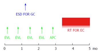
Figure 2 Treatment strategy.
Repeated biweekly endoscopic variceal ligation therapy was performed in advance before radiation therapy for the esophageal cancer. The gastric cancer was resected by endoscopic submucosal dissection during the same period of time.
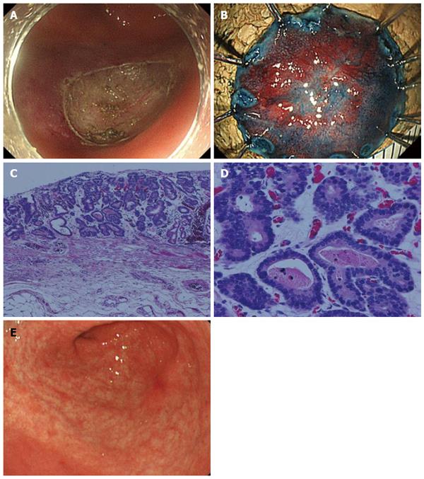
Figure 3 Gastric endoscopic submucosal dissection.
A, B: Successful complete en bloc resection by endoscopic submucosal dissection (ESD) was performed. The operation time was 63 min, and the resected specimen was 22 mm in diameter; C, D: On histology, the lesion was differentiated type mucosal cancer 14 mm in size, and completely resected (magnification: C × 40, D × 200); E: Two months after ESD, the post ESD ulcer was healed and became a red scar.
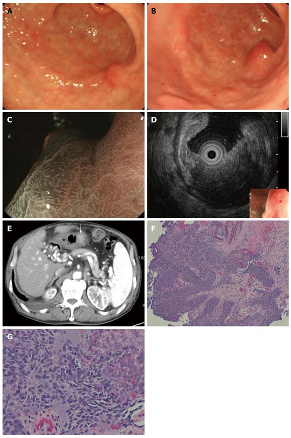
Figure 4 Follow-up esophagogastroduodenoscopy after endoscopic submucosal dissection.
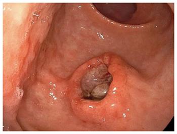
A: A follow-up esophagogastroduodenoscopy (EGD) 7 mo after endoscopic submucosal dissection (ESD) showed a slight elevation on the post ESD scar, but biopsy specimens did not show any malignancy; B: Two more months later, EGD showed a submucosal tumor-like lesion with ulceration 15 mm in size, located right under the post ESD scar; C: Magnifying endoscopy with narrow band imaging showed that the base of the elevation was covered with normal mucosa; D: EUS findings indicated that the tumor mainly involved the submucosal layer; E: The tumor was also recognized as well-demarcated, early enhanced mass on CT; F, G: Biopsy specimens from the edge of the ulcer on the top of the elevated lesion revealed moderately differentiated SCC, which resembled the EC (magnification: F × 40, G × 200).
Figure 5 The tumor seemed to be Borrmann type 2 a year after the endoscopic submucosal dissection.
- Citation: Asai S, Takeshita K, Kano Y, Nakao E, Ichinona T, Fujimoto N, Akamine E, Mori T, Ogawa A. Implantation of esophageal cancer onto post-dissection ulcer after gastric endoscopic submucosal dissection. World J Gastroenterol 2016; 22(9): 2855-2860
- URL: https://www.wjgnet.com/1007-9327/full/v22/i9/2855.htm
- DOI: https://dx.doi.org/10.3748/wjg.v22.i9.2855


