Copyright
©The Author(s) 2016.
World J Gastroenterol. Jun 28, 2016; 22(24): 5512-5519
Published online Jun 28, 2016. doi: 10.3748/wjg.v22.i24.5512
Published online Jun 28, 2016. doi: 10.3748/wjg.v22.i24.5512
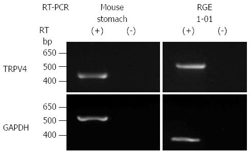
Figure 1 TRPV4 mRNA expression in mouse stomach and RGE1-01 cells.
TRPV4 and GAPDH mRNA levels were examined with (+) and without (-) RT reaction. The expected sizes of the amplified fragments for TRPV4 and GAPDH were 404 and 545 bp for mouse and 524 and 268 bp for rat, respectively. TRPV4 mRNA was detected in mouse stomach and RGE1-01 cells (RGE1-01: normal rat gastric epithelial cell line). Band positions differed due to the use of different primers.
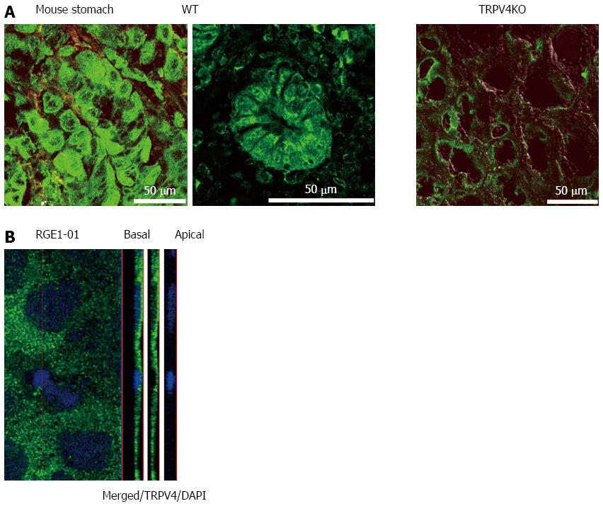
Figure 2 TRPV4 protein expression in mouse stomach and RGE1-01 cells.
A: TRPV4 expression was homogeneously observed in WT but not TRPV4KO mouse gastric epithelium. Bars indicate 50 μm; B: Z-stack image of RGE1-01 cells demonstrated apical TRPV4 expression.
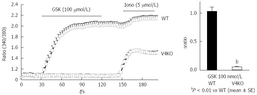
Figure 3 TRPV4-mediated increases in cytosolic Ca2+ ([Ca2+]i) in mouse primary gastric epithelial cells.
A: [Ca2+]i changes (340/380 ratio) in response to the TRPV4 specific agonist GSK1016790A (GSK, 100 nmol/L) in WT or TRPV4KO (V4KO) primary gastric epithelial cells (mean ± SEM). Ionomycin (iono, 5 μmol/L) was used as a positive control. Bars indicate the period of chemical application; B: GSK significantly increased [Ca2+]i in WT cells (means ± SD; 1.03 ± 0.07, n = 20) compared to TRPV4KO cells (0.05 ± 0.01, n = 20) (bP < 0.01 vs WT).
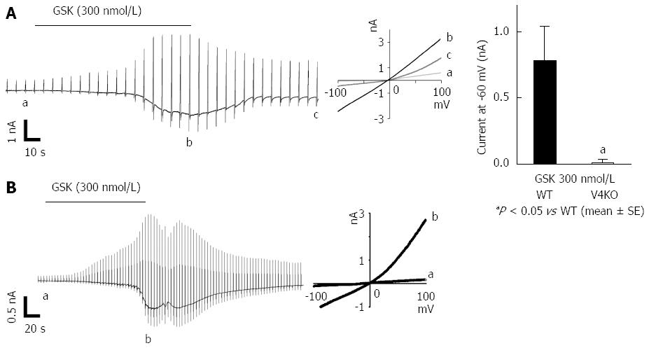
Figure 4 TRPV4-mediated current responses in mouse primary gastric epithelial cells and RGE1-01 cells.
A: GSK (300 nmol/L) evoked inward current responses in WT primary gastric epithelial cells. Currents in response to ramp-pulses at points a, b and c (left in panel B) are shown (middle), with a strongly outwardly rectifying current-voltage relationship. Significantly larger inward currents at -60 mV were obtained from WT cells (means ± SEM; 0.76 ± 0.27 nA, n = 5) than in TRPV4KO cells (0.01 ± 0.00 nA, n = 5) (aP < 0.05 vs WT). B: Similar current responses were also obtained in RGE1-01 cells.
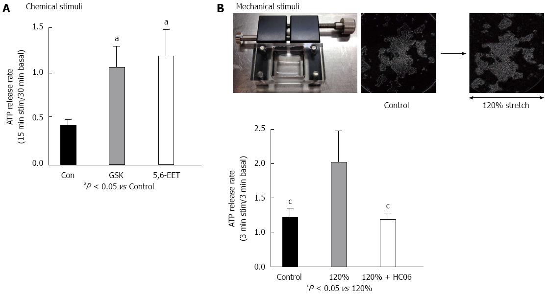
Figure 5 TRPV4 activator-induced ATP release in the RGE1-01 cells.
A: GSK1016790A (GSK, 100 nmol/L) or 5,6-EET (500 nmol/L) induced significantly higher ATP release in RGE1-01 cells (aP < 0.05 vs Control). B: Mechanical stimuli were quantitatively applied with a stretch apparatus. Microscopy images demonstrated that cells were stretched laterally without detachment. A 120% stretch induced significantly higher amounts of ATP release from RGE1-01 cells [cP < 0.05 vs 0% stretch (control)] that could be inhibited by pre-treatment with specific TRPV4 antagonist HC 067047 (1 μmol/L). TRPV4: Transient receptor potential vanilloid 4.
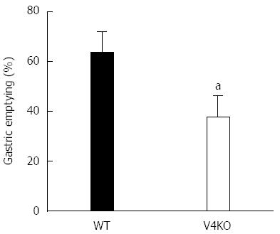
Figure 6 Delayed gastric empting in TRPV4 knockout mice.
Gastric emptying rates in vivo in TRPV4KO mice were significantly delayed relative to WT mice (aP < 0.05, vs WT, n = 7-9).
- Citation: Mihara H, Suzuki N, Boudaka AA, Muhammad JS, Tominaga M, Tabuchi Y, Sugiyama T. Transient receptor potential vanilloid 4-dependent calcium influx and ATP release in mouse and rat gastric epithelia. World J Gastroenterol 2016; 22(24): 5512-5519
- URL: https://www.wjgnet.com/1007-9327/full/v22/i24/5512.htm
- DOI: https://dx.doi.org/10.3748/wjg.v22.i24.5512


