Copyright
©The Author(s) 2016.
World J Gastroenterol. May 21, 2016; 22(19): 4662-4672
Published online May 21, 2016. doi: 10.3748/wjg.v22.i19.4662
Published online May 21, 2016. doi: 10.3748/wjg.v22.i19.4662
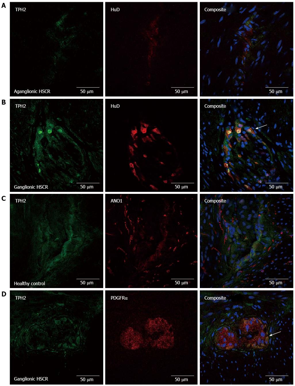
Figure 1 Confocal micrograph series demonstrating pattern of tryptophan hydroxylase-2 expression in the myenteric plexus of the colon.
There is almost no HuD or TPH2 immunofluorescence seen in aganglionic colon in HSCR in (A), while there is clear co-expression (arrow head) in the nerve cell bodies in (B). While TPH2 and anoctamin-1 (ANO1) are not co-expressed in (C), the interstitial cells of Cajal (ICC) fibers form a dense network around TPH2-immuno-positive ganglia. TPH2 was co-expressed with PDGFRα cell bodies in the myenteric plexus, seen in (D). TPH2: Tryptophan hydroxylase-2; HSCR: Hirschsprung’s disease.
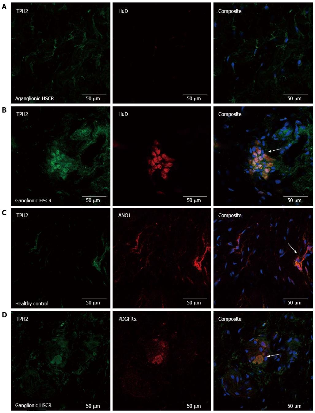
Figure 2 Confocal micrograph series demonstrating tryptophan hydroxylase-2 expression pattern in the submucosal plexus.
Again, no HuD expression is seen in the aganglionic bowel, although there are some TPH2 immuno-positive fibres seen in (A). In ganglionic bowel in HSCR (B), TPH2 co-expressed with HuD (white arrow) in ganglion cells. There was co-expression of TPH2 with anoctamin (ANO)-positive submucosal interstitial cells of Cajals (ICCs) and PDGFRα+ cells, seen in (C) and (D) respectively (white arrow). TPH2: Tryptophan hydroxylase-2; HSCR: Hirschsprung’s disease.
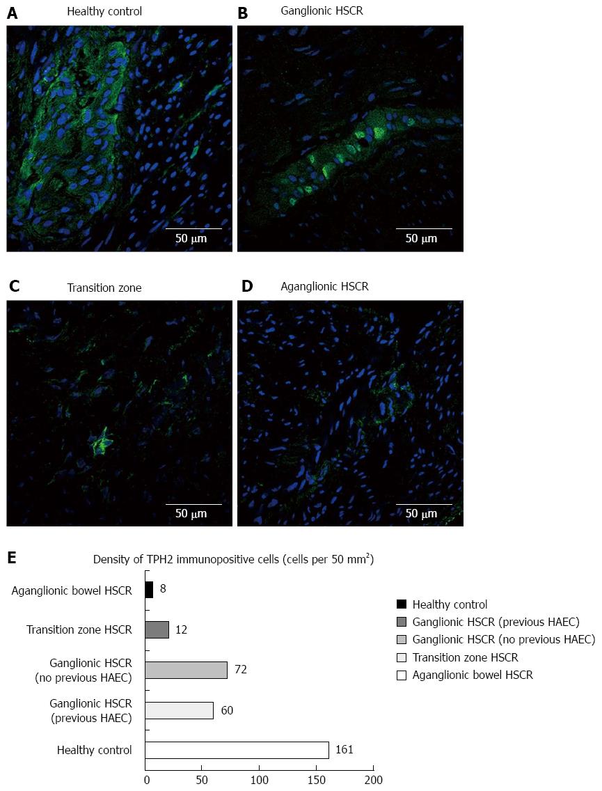
Figure 3 Double-labelled immunofluorescence.
Series (A) to (D) demonstrate the incremental reduction in tryptophan hydroxylase-2 immuno-positive cells in the myenteric plexus from healthy control, to ganglionic bowel in HSCR through to aganglionic bowel. The bar graph in (E) demonstrates the mean cell counts of TPH2 immuno-positive cells, with a reduction seen particularly in transition zone and aganglionic bowel, and slight variation seen in the ganglionic bowel of those with pre-operative HAEC vs those who did not. TPH2: Tryptophan hydroxylase-2; HSCR: Hirschsprung’s disease; HAEC: Hirschsprung’s-associated enterocolitis.
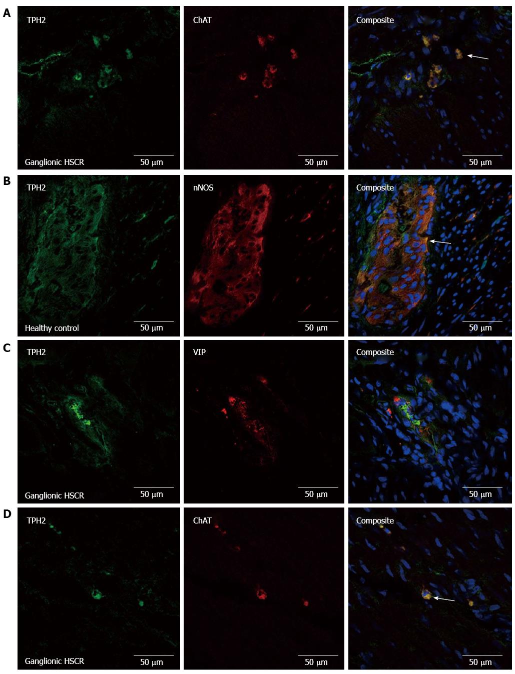
Figure 4 Confocal micrograph series.
Confocal micrograph series demonstrating the cholinergic nature of TPH2 immuno-positive neurons in the myenteric plexus (A), with ChAT-TPH2 co-expression (white arrow). There was also co-expression of nNOS and TPH2 in the myenteric plexus (B) (white arrow) and submucosal plexus (not shown). VIPergic neurons did not express TPH2 in the myenteric plexus (C). Image (D) shows ChAT immune-positive cholinergic nerve fibres in the circular muscle layer, co-expressing TPH2. TPH2: Tryptophan hydroxylase-2.
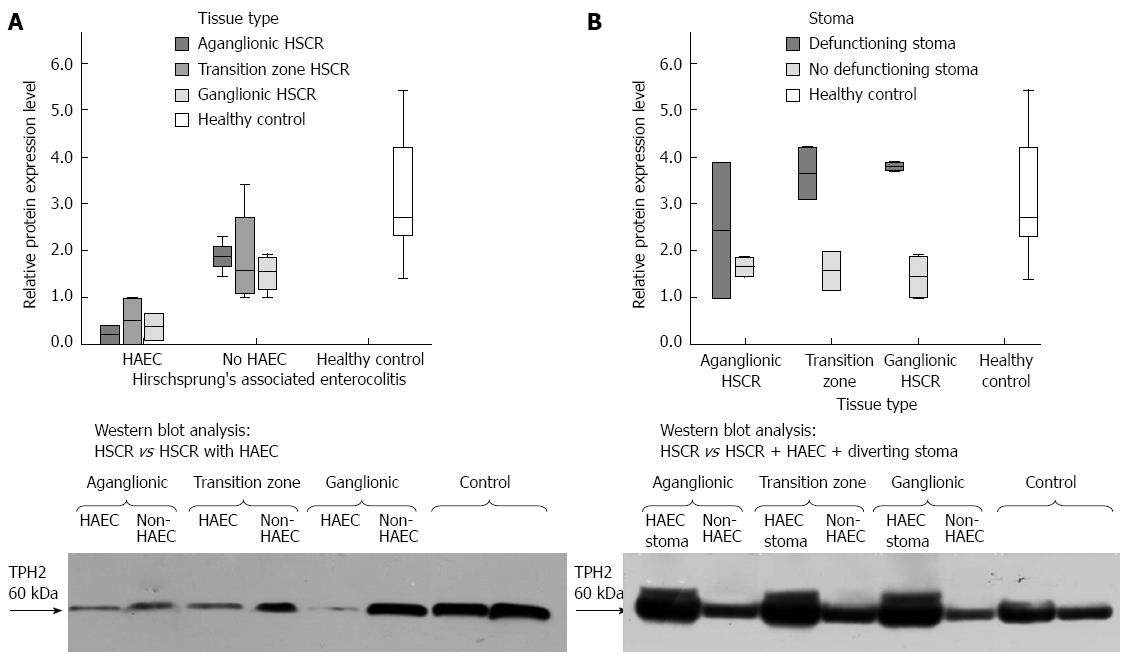
Figure 5 Western blot analysis.
Western blot analysis is seen in (A) showing reduced expression of TPH2 in the colon of children with HSCR complicated by HAEC, who were managed non-operatively prior to pull-through surgery. The reduction in expression is seen across the aganglionic and ganglionic bowel of these patients. Image (B) shows the impact of diverting stoma formation on TPH2 expression in children with HSCR and pre-operative HAEC, with increased expression seen when compared to children without a history of HAEC and no stoma. In both (A) and (B), protein expression has been normalized against the loading control, GAPDH (36kDa). TPH2: Tryptophan hydroxylase-2; HSCR: Hirschsprung’s disease; HAEC: Hirschsprung’s-associated enterocolitis.
- Citation: Coyle D, Murphy JM, Doyle B, O’Donnell AM, Gillick J, Puri P. Altered tryptophan hydroxylase 2 expression in enteric serotonergic nerves in Hirschsprung’s-associated enterocolitis. World J Gastroenterol 2016; 22(19): 4662-4672
- URL: https://www.wjgnet.com/1007-9327/full/v22/i19/4662.htm
- DOI: https://dx.doi.org/10.3748/wjg.v22.i19.4662


