Copyright
©The Author(s) 2016.
World J Gastroenterol. Mar 21, 2016; 22(11): 3242-3251
Published online Mar 21, 2016. doi: 10.3748/wjg.v22.i11.3242
Published online Mar 21, 2016. doi: 10.3748/wjg.v22.i11.3242
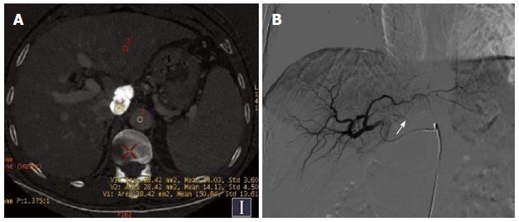
Figure 1 Hepatocellular cancer in the caudate lobe after transcatheter arterial chemoembolization.
A: Iodine (water) based image. The effective iodine content of the defect of iodine deposition (154.10 ± 8.07) was significantly higher than that for normal liver parenchyma and the aorta (67.36 ± 4.87), revealing no residual tumor; B: Digital subtraction angiography image revealed no tumor vessels or stain, and obvious iodized oil deposition was visible (arrow).
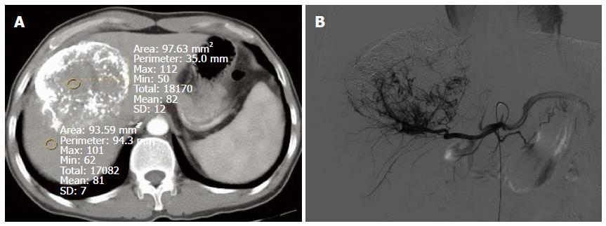
Figure 2 Giant hepatocellular cancer in the lateral borders of the left and right liver lobes after transcatheter arterial chemoembolization.
A: The computed tomography (CT) value of the defect of iodine deposition was comparable to that of normal liver parenchyma in conventional CT image, and there was no obvious enhancement; B: Digital subtraction angiography revealed enlarged tumor blood vessels and tumor stain in the defect of iodine deposition.
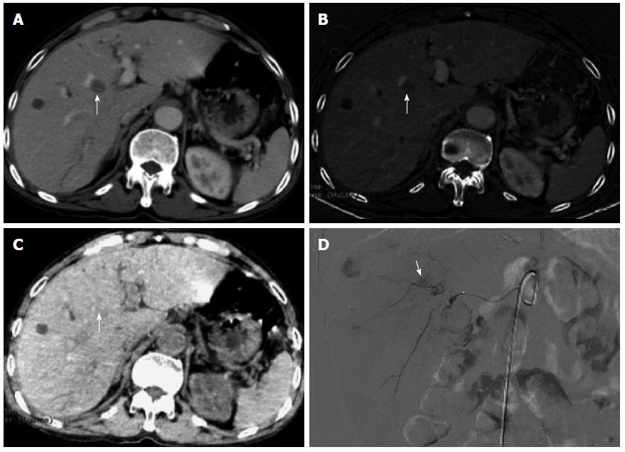
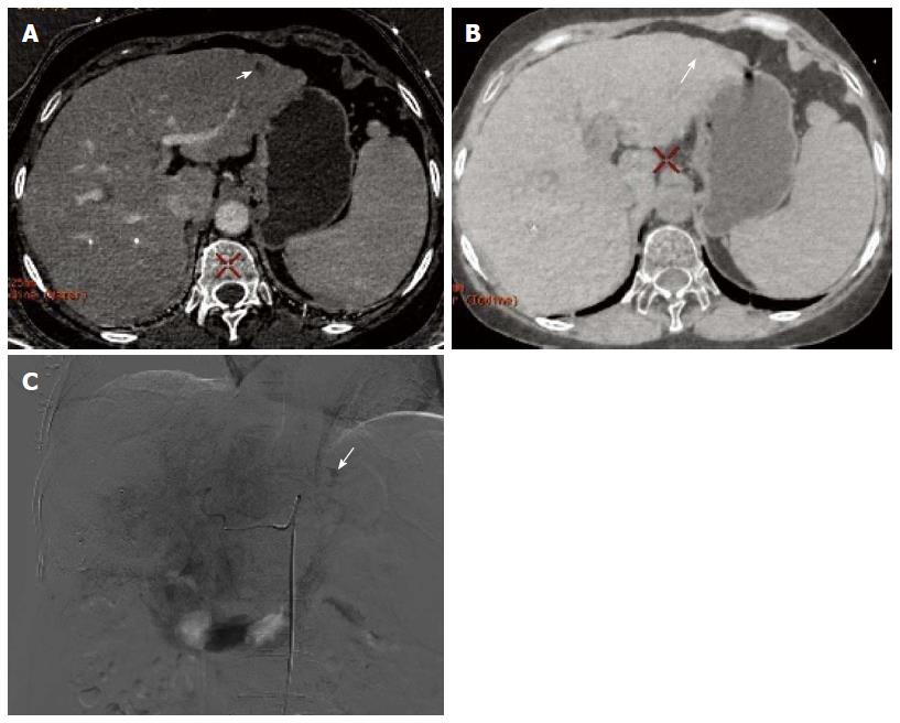
Figure 3 Metastasis to the medial segment of the left lobe of the liver after transcatheter arterial chemoembolization for primary hepatocellular cancer.
A: Conventional computed tomography revealed that the left lobe metastasis had no obvious enhancement; B: Iodine (water) based image showed a small amount of contrast medium entry to the left lobe metastasis, suggesting slight enhancement; C: Iodine (water) based image showed equidensity of the left lobe metastasis; D: Digital subtraction angiography confirmed tumor stain in the left lobe metastasis in parenchymal phase (arrow).
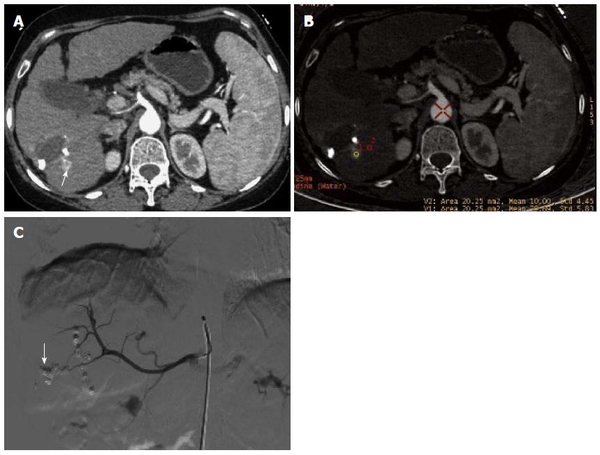
Figure 4 Recurrence of hepatocellular cancer in the right lobe after transcatheter arterial chemoembolization.
A: Sixty-eight keV monochromatic image showed obvious enlargement and enhancement of recurrent lesion in tumor edge (arrow); B: The effective iodine content of the recurrent lesion (29.89 ± 5.83) was significantly higher than that for normal liver parenchyma (10.0 ± 4.45), suggesting obvious enhancement and entry of contrast medium to the lesion; C: Digital subtraction angiography confirmed tumor stain in the recurrent lesion in parenchymal phase (arrow).
Figure 5 Same case as in Figure 4.
A: Iodine (water) based image revealed no entry of contrast medium to the lateral segment of the left lobe, which showed hypodensity (arrow); B: Iodine (water) based image showed slight hypodensity (arrow); C: Digital subtraction angiography confirmed tumor stain in the left lobe metastasis in parenchymal phase (arrow).
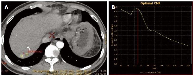
Figure 6 Metastasis to the right lobe after transcatheter arterial chemoembolization for hepatocellular cancer in the right lobe.
A: Placement of regions of interest in the metastatic lesion and surrounding normal parenchyma, respectively; B: Carrier-to-noise ratio curve. The optimal monochromatic images were at 60-70 keV.
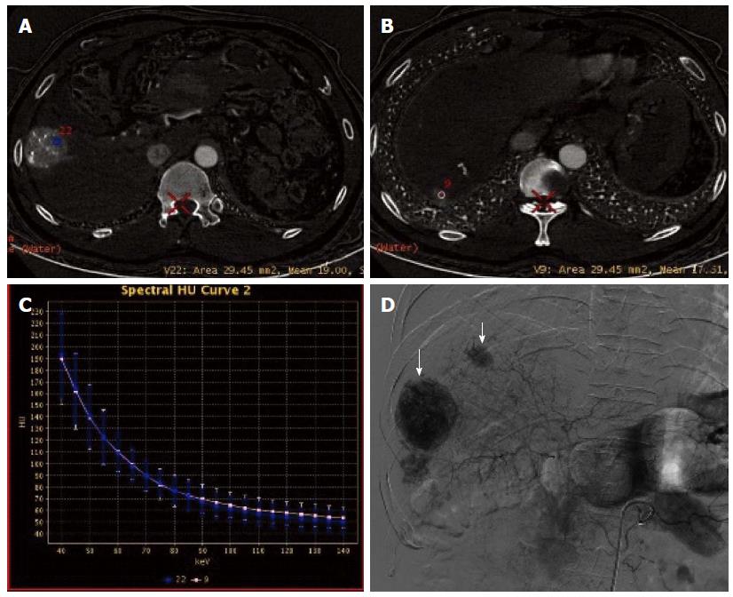
Figure 7 Same case as in Figure 6.
A: Placement of a region of interest in the primary lesion; B: Placement of a region of interest in the lesion in the right lobe; C: The spectral curves for the two lesions were roughly same, suggesting tumor homogeneity; D: Digital subtraction angiography confirmed multiple tumor stains (arrow).
- Citation: Liu QY, He CD, Zhou Y, Huang D, Lin H, Wang Z, Wang D, Wang JQ, Liao LP. Application of gemstone spectral imaging for efficacy evaluation in hepatocellular carcinoma after transarterial chemoembolization. World J Gastroenterol 2016; 22(11): 3242-3251
- URL: https://www.wjgnet.com/1007-9327/full/v22/i11/3242.htm
- DOI: https://dx.doi.org/10.3748/wjg.v22.i11.3242


