Copyright
©The Author(s) 2016.
World J Gastroenterol. Mar 21, 2016; 22(11): 3227-3233
Published online Mar 21, 2016. doi: 10.3748/wjg.v22.i11.3227
Published online Mar 21, 2016. doi: 10.3748/wjg.v22.i11.3227
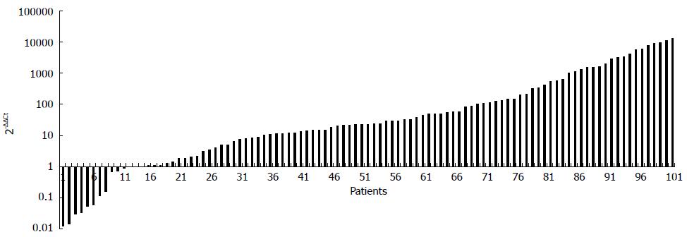
Figure 1 Fold change of Fusobacterium nucleatum abundance in colorectal cancer vs matched normal tissues (n = 101).
2-ΔΔCT represented the fold change of Fusobacterium nucleatum load in cancer tissue over matched normal tissues. The numbers in Y-axis indicated the 2-ΔΔCT value of each patient.
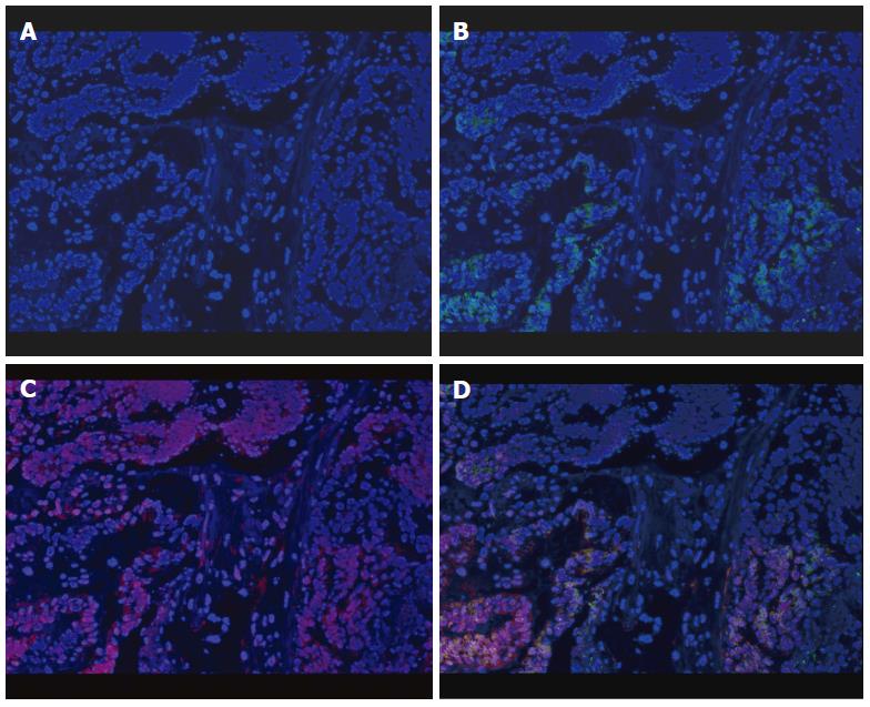
Figure 2 Enrichment of Fusobacterium nucleatum in colorectal cancer detected by FISH.
Graph A: Colorectal cancer (CRC) specimen stained with DAPI. Cell nuclei were stained in blue; B: CRC specimen stained with both DAPI and universal bacterial probe (EUB338). Bacterial conserved regions were stained in green; C: CRC specimen stained with both DAPI and Fusobacterium specific probe (FUSO). The Fusobacterium nucleatum (F. nucleatum) specific regions were stained in red; D: CRC specimen triply stained with DAPI, EUSO and EUB338. Cell nuclei (in blue), bacterial conserved regions (in green) and F. nucleatum specific regions (in red) were clearly visible (all × 400).
- Citation: Li YY, Ge QX, Cao J, Zhou YJ, Du YL, Shen B, Wan YJY, Nie YQ. Association of Fusobacterium nucleatum infection with colorectal cancer in Chinese patients. World J Gastroenterol 2016; 22(11): 3227-3233
- URL: https://www.wjgnet.com/1007-9327/full/v22/i11/3227.htm
- DOI: https://dx.doi.org/10.3748/wjg.v22.i11.3227


