Copyright
©The Author(s) 2015.
World J Gastroenterol. Feb 21, 2015; 21(7): 2191-2198
Published online Feb 21, 2015. doi: 10.3748/wjg.v21.i7.2191
Published online Feb 21, 2015. doi: 10.3748/wjg.v21.i7.2191
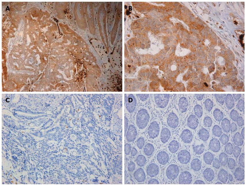
Figure 1 Hypoxia-inducible factor-1α expression patterns.
A: A sample with strong cytoplasmic staining for hypoxia-inducible factor-1α (HIF-1α) in the advancing tumor border is shown; interstitial cells are positive for HIF-1α (× 200); B: Cancer tissue with positive immunostaining for HIF-1α (× 400); C: Cancer tissue with negative immunostaining for HIF-1α (× 200); D: HIF-1α is not expressed in normal tissue (× 200).
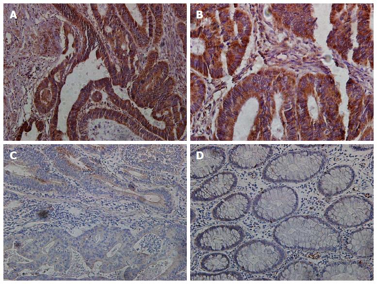
Figure 2 Expression patterns of semaphorin 4D.
A: A sample with strong cytoplasmic staining for semaphorin 4D (Sema4D) in the advancing tumor border is shown, with interstitial cells being positive for Sema4D (× 200); B: Cancer tissue with positive immunostaining for Sema4D (× 400); C: Cancer tissue with negative immunostaining for Sema4D (× 200); D: Sema4D is not expressed in normal tissue (× 200).
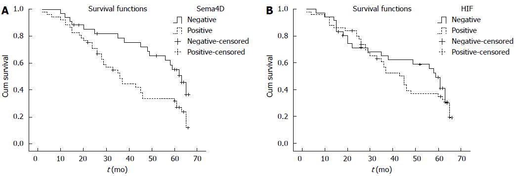
Figure 3 Cumulative survival curves.
Cumulative survival curves were stratified by semaphorin 4D (Sema4D) and hypoxia-inducible factor-1α (HIF-1α) expression in cancer cells. The survival curves were analyzed by Kaplan-Meier analysis and log-rank tests. A: Colorectal carcinoma patients with positive Sema4D expression exhibited shorter overall survival compared with patients with negative expression (P = 0.009); B: Colorectal carcinoma patients with positive HIF-1α expression exhibited shorter overall survival compared with those with negative expression (P = 0.497). Neither value reached statistical significance.
- Citation: Wang JS, Jing CQ, Shan KS, Chen YZ, Guo XB, Cao ZX, Mu LJ, Peng LP, Zhou ML, Li LP. Semaphorin 4D and hypoxia-inducible factor-1α overexpression is related to prognosis in colorectal carcinoma. World J Gastroenterol 2015; 21(7): 2191-2198
- URL: https://www.wjgnet.com/1007-9327/full/v21/i7/2191.htm
- DOI: https://dx.doi.org/10.3748/wjg.v21.i7.2191


