Copyright
©The Author(s) 2015.
World J Gastroenterol. Feb 21, 2015; 21(7): 2169-2177
Published online Feb 21, 2015. doi: 10.3748/wjg.v21.i7.2169
Published online Feb 21, 2015. doi: 10.3748/wjg.v21.i7.2169
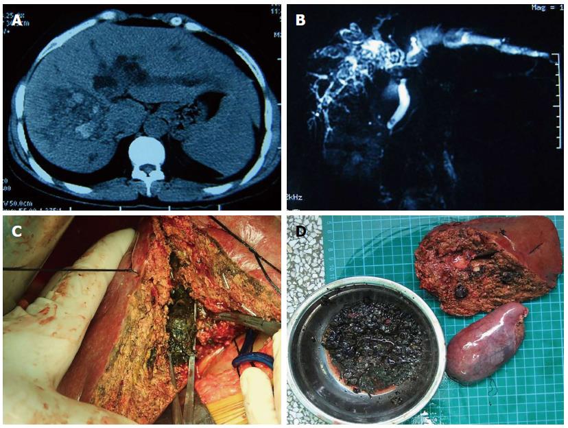
Figure 1 Primary type of pathological evolution-based classification for hepatolithiasis.
A: Computed tomography showed that calculi were located within the right posterior lobe of the liver and dilation of the biliary tract within the liver; B: Magnetic resonance cholangiopancreatography showed dilation of the biliary tract and a negative calculi signal within the dilated tract; C: Operative imaging showed that a large amount of hepatolithiasis was removed surgically; D: Postoperative imaging showed calculi (in kidney basin) and specimens including gallbladder and posterior hepatic lobe, which were removed surgically.
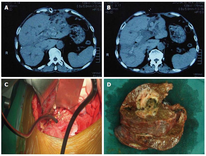
Figure 2 Inflammatory type of pathological evolution-based classification for hepatolithiasis.
A: Computed tomography (CT) showed postoperative changes in the liver after three operations on the biliary tract; B: CT showed dilation of the biliary tract and calculi within the dilated tract in the same patient; C: Operative imaging showed intraoperative choledochoscopy aided intraoperative stone extraction; D: Postoperative imaging showed left lateral lobe of the liver with calculi inside, which were removed during the operation.
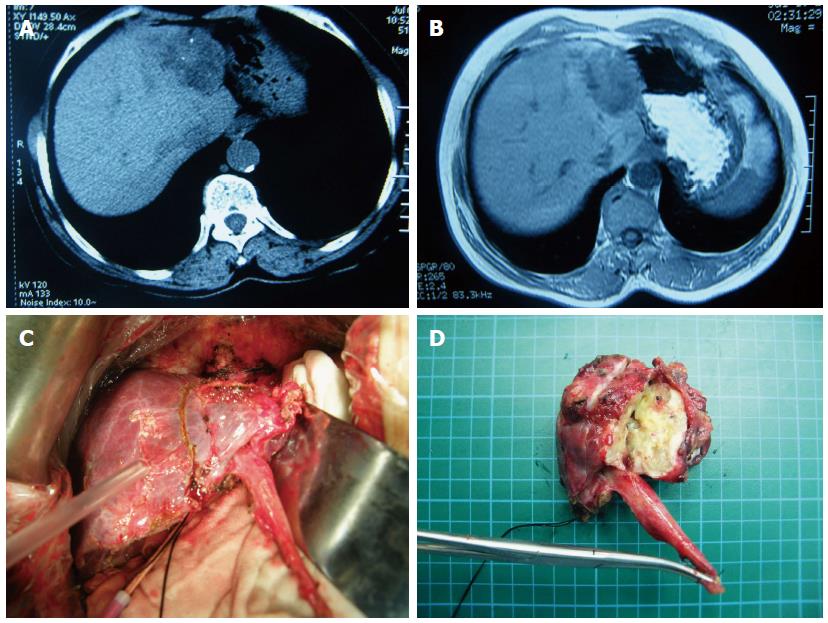
Figure 3 Mass-forming type of pathological evolution-based classification for hepatolithiasis.
A: Computed tomography showed mass that formed in the left lobe of the liver with positive calculi signal inside the mass; B: Magnetic resonance cholangiopancreatography showed that the same lesion was reflected in the same area in the liver; C: Operative imaging showed that the left lobe was removed intraoperatively; D: Postoperative imaging showed that the resected left lobe contained the mass (postoperative pathology report: mucinous adenocarcinoma).
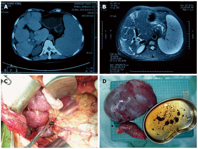
Figure 4 Terminal type of pathological evolution-based classification for hepatolithiasis.
A: Computed tomography showed liver cirrhosis and splenomegaly; B: Magnetic resonance cholangiopancreatography showed liver cirrhosis (schistosomiasis cirrhosis) and splenomegaly; C: Operative imaging showed liver cirrhosis and removal of cholecyst; D: Postoperative imaging showed cholecyst removal, enlarged spleen and extracted calculi (in kidney basin).
- Citation: Liu FB, Yu XJ, Wang GB, Zhao YJ, Xie K, Huang F, Cheng JM, Wu XR, Liang CJ, Geng XP. Preliminary study of a new pathological evolution-based clinical hepatolithiasis classification. World J Gastroenterol 2015; 21(7): 2169-2177
- URL: https://www.wjgnet.com/1007-9327/full/v21/i7/2169.htm
- DOI: https://dx.doi.org/10.3748/wjg.v21.i7.2169


