Copyright
©The Author(s) 2015.
World J Gastroenterol. Feb 21, 2015; 21(7): 2140-2146
Published online Feb 21, 2015. doi: 10.3748/wjg.v21.i7.2140
Published online Feb 21, 2015. doi: 10.3748/wjg.v21.i7.2140
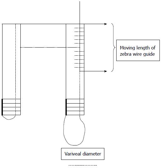
Figure 1 Diagram describing the principle of this endoscopic measuring scale.
A: Closing loop of stylet; B: Opening loop of stylet.
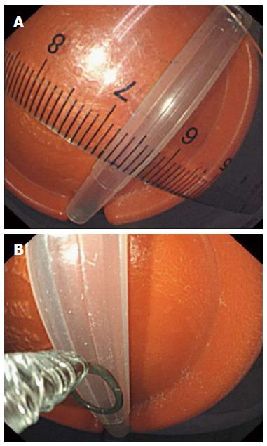
Figure 2 Two different methods of measurement; the diameter of the simulative varices was 0.
6 and 0.5 cm, respectively. A: Ruler measuring diameter 0.6 cm; B: Endoscopic measuring scale diameter 0.5 cm.
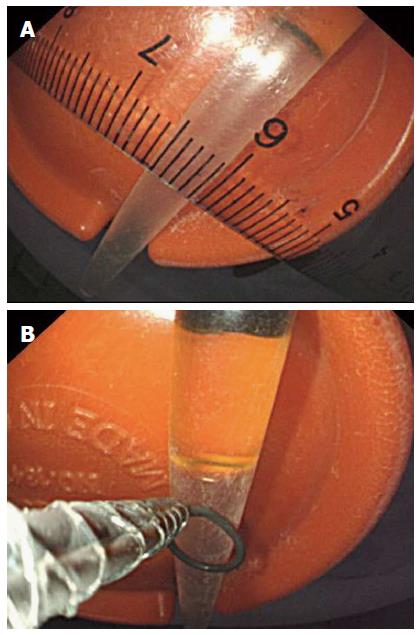
Figure 3 Two different methods of measurement; the diameter of the simulative varices was 0.
45 and 0.5 cm, respectively. A: Ruler measuring diameter 0.45 cm; B: Endoscopic measuring scale diameter 0.5 cm.
- Citation: Li ZQ, Linghu EQ, Hu M, Wang XD, Wang HB, Meng JY, Du H. Endoscopic measurement of variceal diameter. World J Gastroenterol 2015; 21(7): 2140-2146
- URL: https://www.wjgnet.com/1007-9327/full/v21/i7/2140.htm
- DOI: https://dx.doi.org/10.3748/wjg.v21.i7.2140


