Copyright
©The Author(s) 2015.
World J Gastroenterol. Nov 14, 2015; 21(42): 11954-11963
Published online Nov 14, 2015. doi: 10.3748/wjg.v21.i42.11954
Published online Nov 14, 2015. doi: 10.3748/wjg.v21.i42.11954
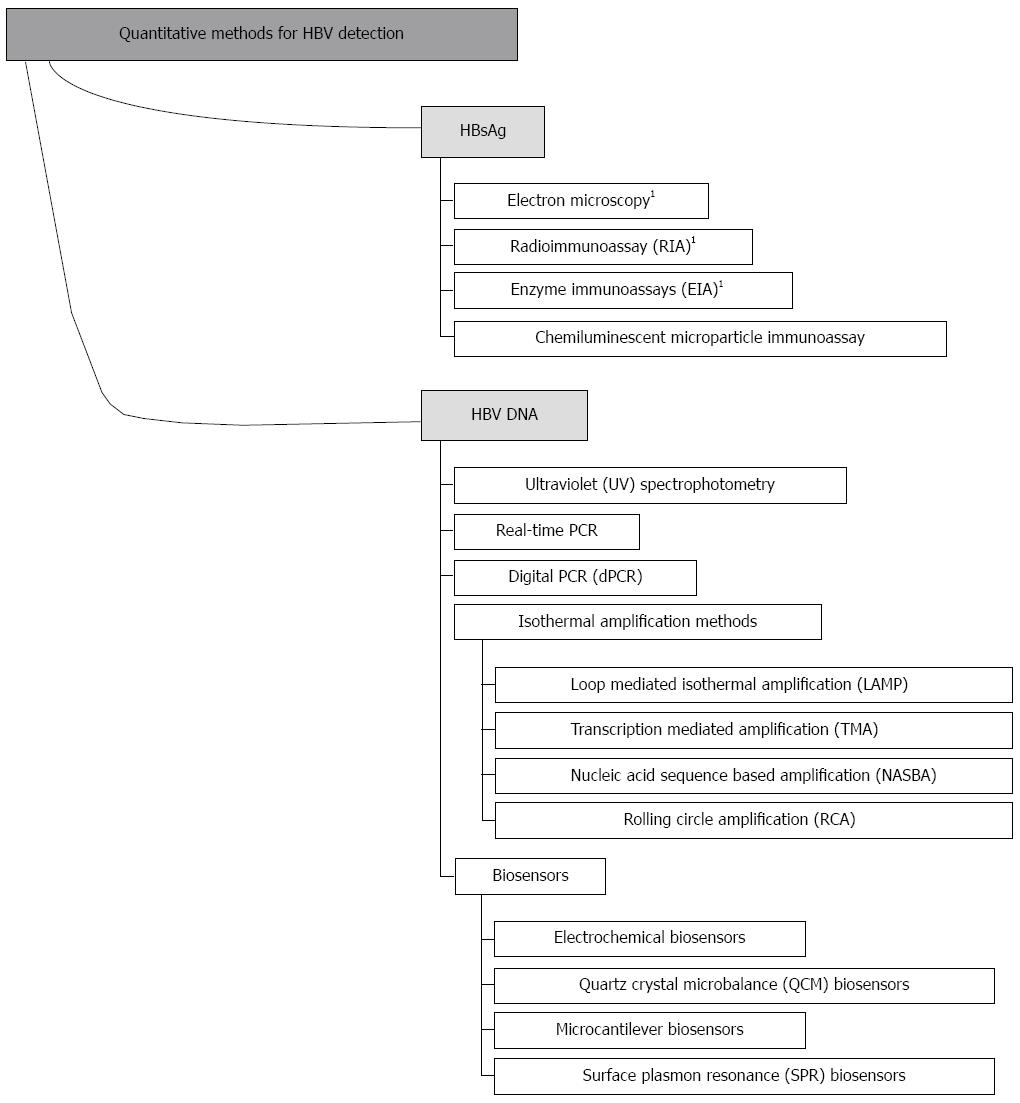
Figure 1 Quantitative methods for hepatitis B virus detection.
1These methods are qualitative or semi-quantitative for HBsAg. HBsAg: Hepatitis B surface antigen.
Figure 2 Reaction mechanism of real-time polymerase chain reaction based on TaqMan probe technology.
TaqMan probe is an oligo-nucleotide probe that has a fluorescent reporter at the 5’ end and a quencher attached to the 3’ end. Once hybridized to the target sequence during annealing, TaqMan probe is cleaved by DNA polymerase, which separates the fluorescent reporter from the quencher. Once they are separated, the signal is emitted and detected in the real-time machine. The intensity of fluorescence is proportional to the amount of PCR product produced. FRET: Fluorescent resonance energy transfer.
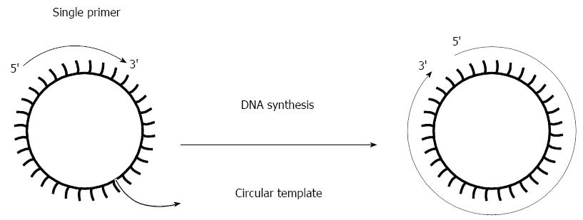
Figure 3 Scheme of the rolling circle amplification reaction.
RCA rapidly synthesizes multiple copies of a single circular template with use of a single primer. RCA: Rolling circle amplification.
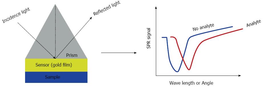
Figure 4 Scheme of the surface plasmon resonance biosensor.
The incidence light causes the changes in wavelength or angle. Changes were detected in real time monitor. SPR: Surface plasmon resonance.
- Citation: Liu YP, Yao CY. Rapid and quantitative detection of hepatitis B virus. World J Gastroenterol 2015; 21(42): 11954-11963
- URL: https://www.wjgnet.com/1007-9327/full/v21/i42/11954.htm
- DOI: https://dx.doi.org/10.3748/wjg.v21.i42.11954


