Copyright
©The Author(s) 2015.
World J Gastroenterol. Jan 28, 2015; 21(4): 1362-1364
Published online Jan 28, 2015. doi: 10.3748/wjg.v21.i4.1362
Published online Jan 28, 2015. doi: 10.3748/wjg.v21.i4.1362
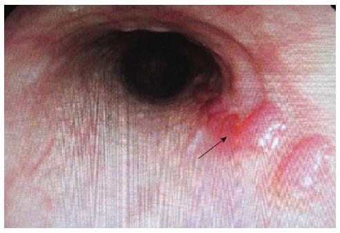
Figure 1 Gastroscope revealed esophageal varices (black arrow).
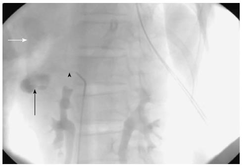
Figure 2 DSA showed a mass of 52 mm × 48 mm (black arrow), the portal vein(white arrow) was highlighted at the arteriaphase(black arrowhead), strongly suggesting the existence of a hepatic arterioportal communication.
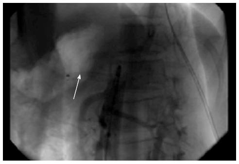
Figure 3 Aortogram revealed no endoleak and no blood flow entering into Portal Vein anymore (arrow).
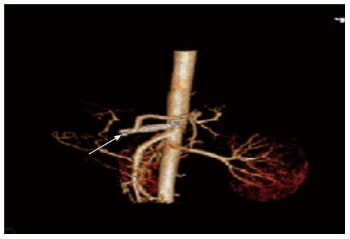
Figure 4 Post-operation recovery of the patient has been uneventful, and no additional episodes of upper gastrointestinal bleeding have been reported by the patient on three subsequent clinic visits during the last six months (arrow).
- Citation: Liu YR, Huang B, Yuan D, Wu ZP, Zhao JC. Unusual case of digestive hemorrhage: Celiac axis-portal vein arteriovenous fistula. World J Gastroenterol 2015; 21(4): 1362-1364
- URL: https://www.wjgnet.com/1007-9327/full/v21/i4/1362.htm
- DOI: https://dx.doi.org/10.3748/wjg.v21.i4.1362


