Copyright
©The Author(s) 2015.
World J Gastroenterol. Sep 28, 2015; 21(36): 10480-10484
Published online Sep 28, 2015. doi: 10.3748/wjg.v21.i36.10480
Published online Sep 28, 2015. doi: 10.3748/wjg.v21.i36.10480
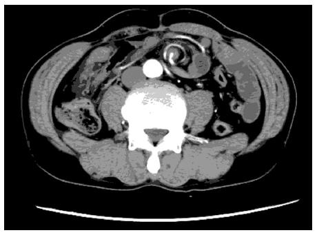
Figure 1 Contrast-enhanced computed tomography scan of the abdomen showing the mesentery and superior mesenteric artery with whirl sign.
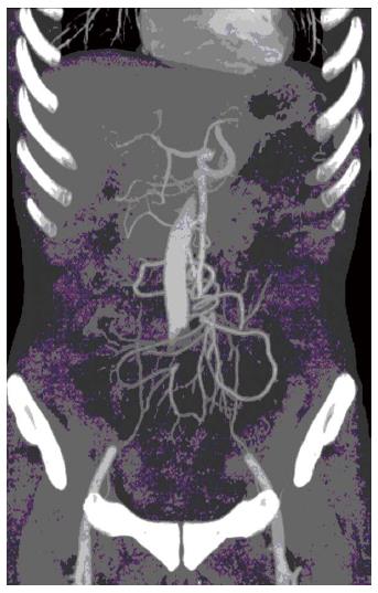
Figure 2 Multidetector computed tomography showing twisted small vessels arising from the distal superior mesenteric artery, with no relevant primary vascular stenosis or occlusion.
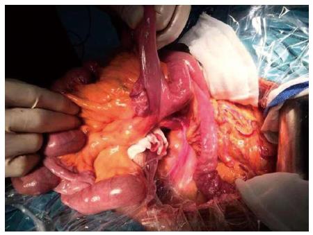
Figure 3 One jejunal diverticulum with a size of 2.
6 cm × 1.8 cm was seen at laparotomy.
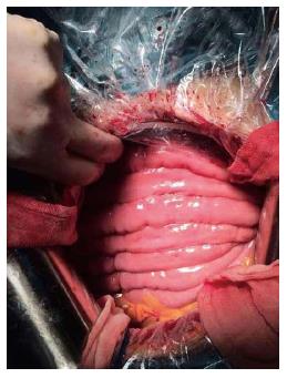
Figure 4 Whole small bowel arrangement to prevent re-torsion was performed at laparotomy.
- Citation: Shen XF, Guan WX, Cao K, Wang H, Du JF. Small bowel volvulus with jejunal diverticulum: Primary or secondary? World J Gastroenterol 2015; 21(36): 10480-10484
- URL: https://www.wjgnet.com/1007-9327/full/v21/i36/10480.htm
- DOI: https://dx.doi.org/10.3748/wjg.v21.i36.10480


