Copyright
©The Author(s) 2015.
World J Gastroenterol. Sep 14, 2015; 21(34): 10049-10053
Published online Sep 14, 2015. doi: 10.3748/wjg.v21.i34.10049
Published online Sep 14, 2015. doi: 10.3748/wjg.v21.i34.10049
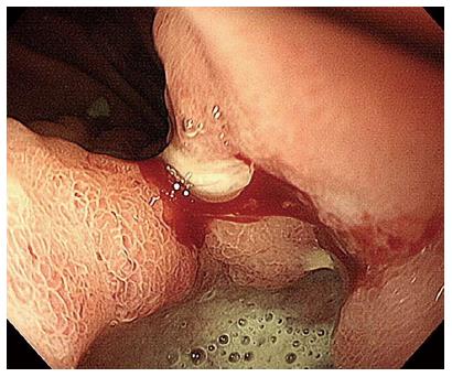
Figure 1 Endoscopic examination showed an 2.
5-cm active gastric malignancy, Borrmann type III.
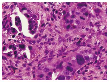
Figure 2 Biopsy showed clusters of adenocarcinoma cells with large hyperchromatic and pleomorphic nuclei.
Mitotic activity is easily identified (arrows). Original magnification × 400.
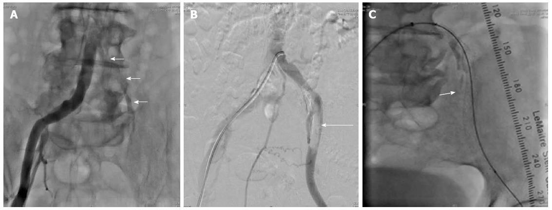
Figure 3 Angiography.
A: Diffuse stenosis of the left common iliac artery without blood flow (arrows); B: thrombi in the left external iliac artery (7.0 × 6.0-cm thrombi, arrow); C: Angiography of the left common iliac artery after thrombus suction and deployment of a complete iliac stent (arrow) from the common iliac artery to the external iliac artery.
- Citation: Chien TL, Rau KM, Chung WJ, Tai WC, Wang SH, Chiu YC, Wu KL, Chou YP, Wu CC, Chen YH, Chuah SK, Gastric Cancer Team. Trousseau’s syndrome in a patient with advanced stage gastric cancer. World J Gastroenterol 2015; 21(34): 10049-10053
- URL: https://www.wjgnet.com/1007-9327/full/v21/i34/10049.htm
- DOI: https://dx.doi.org/10.3748/wjg.v21.i34.10049


