Copyright
©The Author(s) 2015.
World J Gastroenterol. Aug 28, 2015; 21(32): 9534-9543
Published online Aug 28, 2015. doi: 10.3748/wjg.v21.i32.9534
Published online Aug 28, 2015. doi: 10.3748/wjg.v21.i32.9534
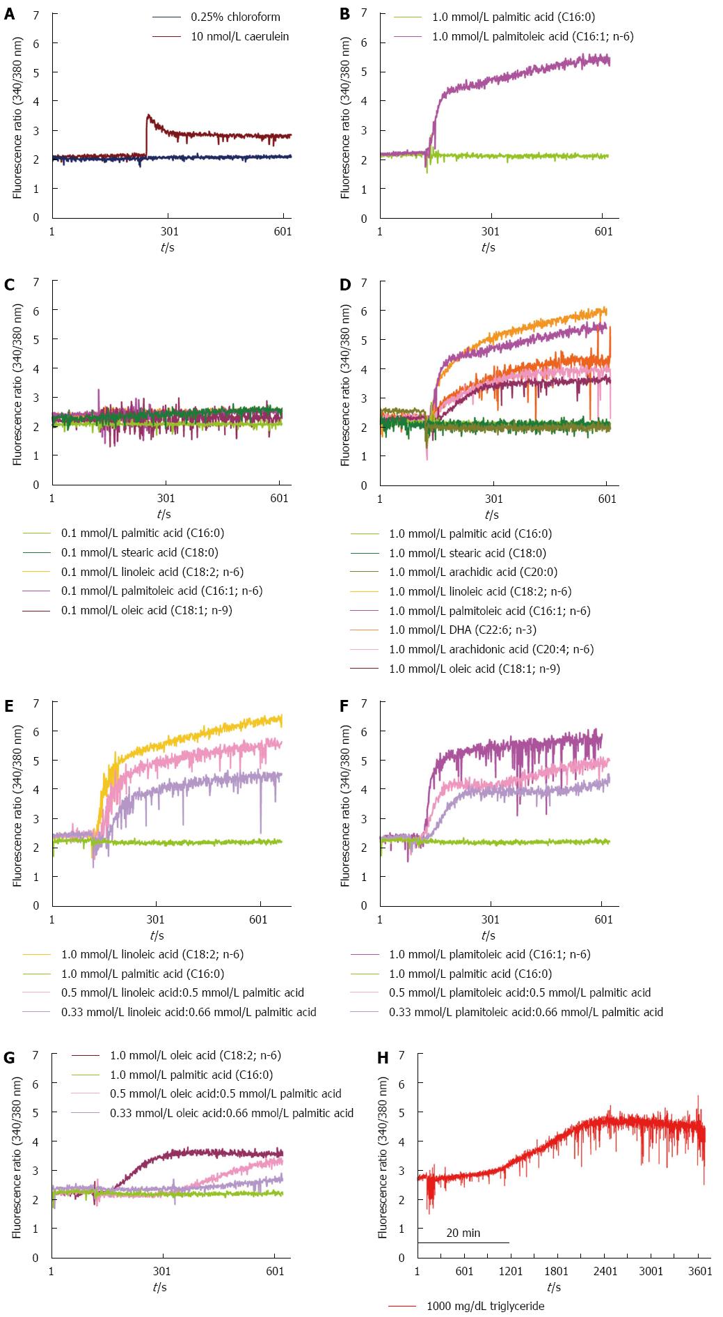
Figure 1 Effects of unsaturated and saturated fatty acids on cytosolic Ca2+ concentrations in acinar cells.
A: A 0.25% chloroform and 10 nmol/L cerulein solution was used as the control; B: A high concentration (1.0 mmol/L) of the unsaturated fatty acid palmitoleic acid, but not of the saturated fatty acid palmitic acid, induced a sustained cytosolic Ca2+ elevation; C: Various unsaturated fatty acids and saturated fatty acids at low concentrations (0.1 mmol/L) did not induce sustained cytosolic Ca2+ elevations; D: Various unsaturated fatty acids at high concentrations (1.0 mmol/L), but not saturated fatty acids, induced sustained cytosolic Ca2+ elevations. The effects of different ratios of unsaturated fatty acids and saturated fatty acids on the elevation of cytosolic Ca2+ concentrations: linoleic acid and palmitic acid (E), palmitoleic acid and palmitic acid (F), and oleic acid and palmitic acid (G); H: High concentrations (1000 mg/dL) of triglycerides did not induce the elevation of cytosolic Ca2+ concentrations.
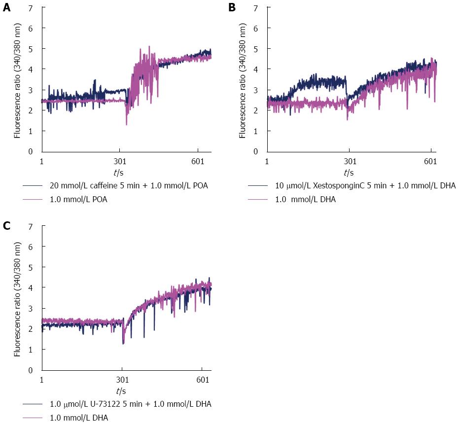
Figure 2 Inhibition of IP3R did not prevent the elevation of cytosolic Ca2+ concentrations in acinar cells induced by unsaturated fatty acids: caffeine (A), xestospongin C (B), and U-73122 (C).
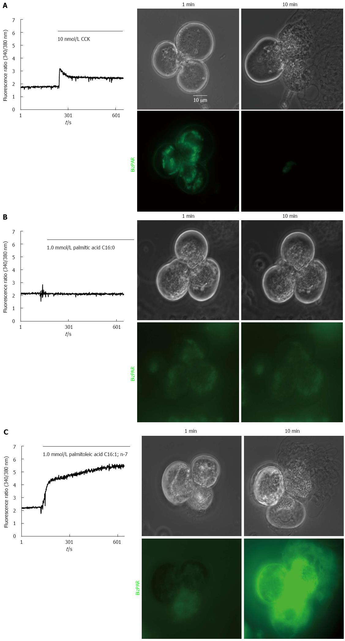
Figure 3 Unsaturated fatty acids at high concentrations (1.
0 mmol/L), but not saturated fatty acids, induced intra-acinar cell trypsin activation and cell damage observed by confocal microscopy: CCK (A), palmitic acid (B), and palmitoleic acid (C).
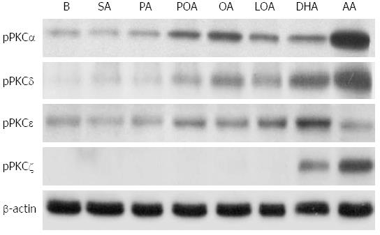
Figure 4 Effects of unsaturated fatty acids at high concentrations (1.
0 mmol/L) on protein kinase C isoform expression in acinar cells as detected by western blot. B: Blank; SA: Stearic acid; PA: Palmitic acid; POA: Palmitoleic acid; OA: Oleic acid; LA: Linoleic acid; DHA: Docosahexaenoic acid; AA: Arachidonic acid.
- Citation: Chang YT, Chang MC, Tung CC, Wei SC, Wong JM. Distinctive roles of unsaturated and saturated fatty acids in hyperlipidemic pancreatitis. World J Gastroenterol 2015; 21(32): 9534-9543
- URL: https://www.wjgnet.com/1007-9327/full/v21/i32/9534.htm
- DOI: https://dx.doi.org/10.3748/wjg.v21.i32.9534


