Copyright
©The Author(s) 2015.
World J Gastroenterol. Jul 21, 2015; 21(27): 8408-8417
Published online Jul 21, 2015. doi: 10.3748/wjg.v21.i27.8408
Published online Jul 21, 2015. doi: 10.3748/wjg.v21.i27.8408
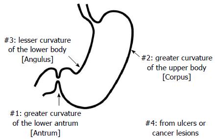
Figure 1 Triple-site biopsy.
Triple-site biopsy specimens were used for the histologic diagnosis of chronic inflammation, neutrophil activity, glandular atrophy, intestinal metaplasia and Helicobacter pylori in the gastric mucosa.
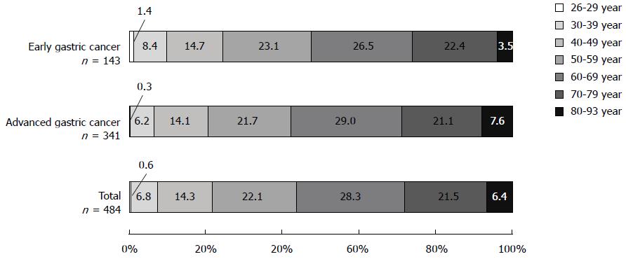
Figure 2 Analysis of gastric cancer in Mongolian patients.
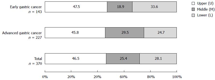
Figure 3 Distribution of gastric cancer in Mongolian patients.
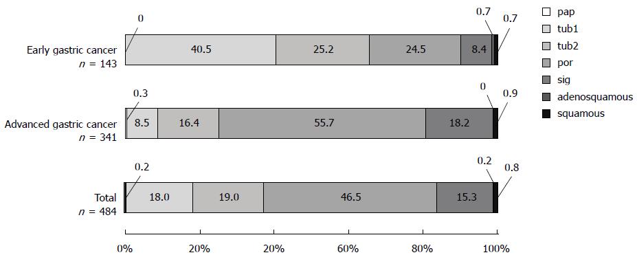
Figure 4 Histologic distribution of gastric cancer in Mongolian patients.
Analysis of the percentages of differentiated adenocarcinoma (papillary adenocarcinoma [pap], well-differentiated type [tub1], and moderately differentiated type [tub2]) and undifferentiated adenocarcinoma (poorly differentiated adenocarcinoma [por] and signet-ring cell carcinoma [sig]).
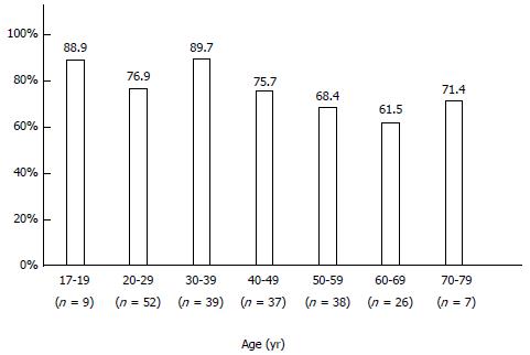
Figure 5 Prevalence of Helicobacter pylori infection in Mongolian patients according to age.
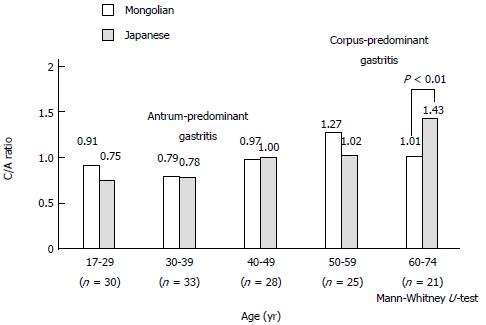
Figure 6 Corpus/antrum activity score ratio in Helicobacter pylori-positive Mongolian and Japanese patients matched by age, sex, and endoscopic diagnostics.
Corpus/antrum (C/A) ratio < 1 indicates antrum-predominant gastritis, whereas ratio > 1 corpus-predominant gastritis.
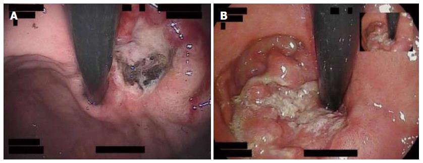
Figure 7 Gastric cancer cases in the upper region.
A: Sixty-seven-year-old-woman with cancer located near the cardia, type 2; B: Eighty-eight-year-old-man with cancer located around the cardia, type 3.
-
Citation: Matsuhisa T, Yamaoka Y, Uchida T, Duger D, Adiyasuren B, Khasag O, Tegshee T, Tsogt-Ochir B. Gastric mucosa in Mongolian and Japanese patients with gastric cancer and
Helicobacter pylori infection. World J Gastroenterol 2015; 21(27): 8408-8417 - URL: https://www.wjgnet.com/1007-9327/full/v21/i27/8408.htm
- DOI: https://dx.doi.org/10.3748/wjg.v21.i27.8408


