Copyright
©The Author(s) 2015.
World J Gastroenterol. Jul 21, 2015; 21(27): 8389-8397
Published online Jul 21, 2015. doi: 10.3748/wjg.v21.i27.8389
Published online Jul 21, 2015. doi: 10.3748/wjg.v21.i27.8389
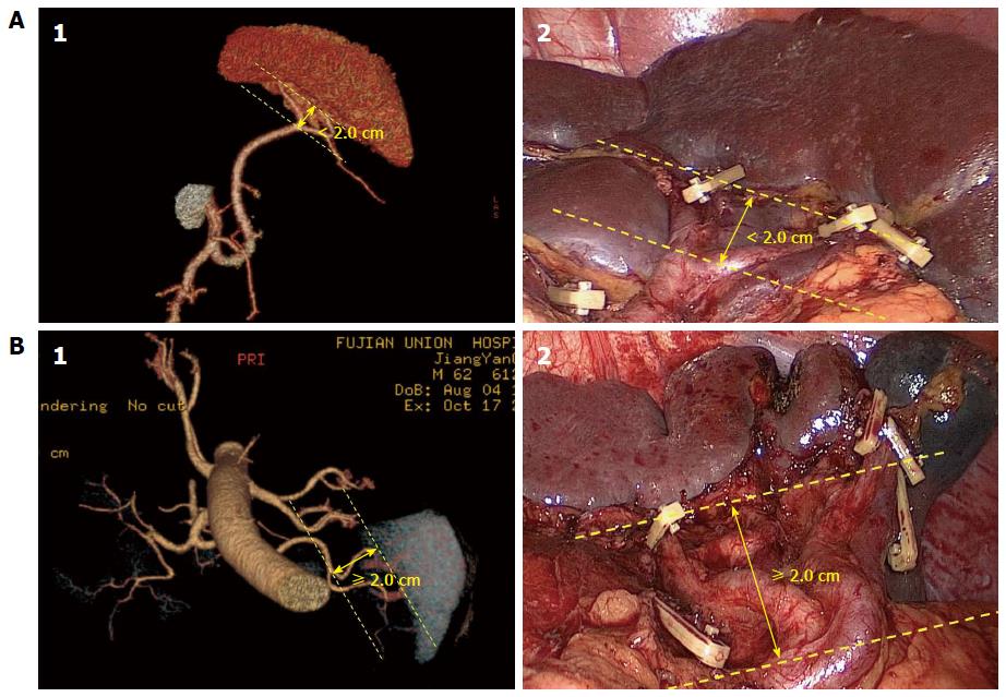
Figure 1 Terminal branches of the splenic artery.
A: The concentrated type SpA (1: Preoperative assessment using 3DCT images; 2: Operative view after the completion of the splenic LN dissection); B: The distributed type SpA (1: Preoperative assessment using 3DCT images; 2: Operative view after the completion of the splenic LN dissection). SpA: Splenic artery; 3DCT: 3-dimensional computer tomography; LNs: Lymph nodes.
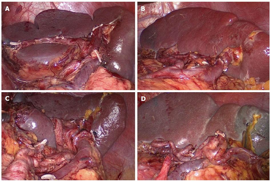
Figure 2 Splenic lobar artery.
A: Single-branch type; B: Two-branched type; C: Three-branched type; D: Multiple-branched.
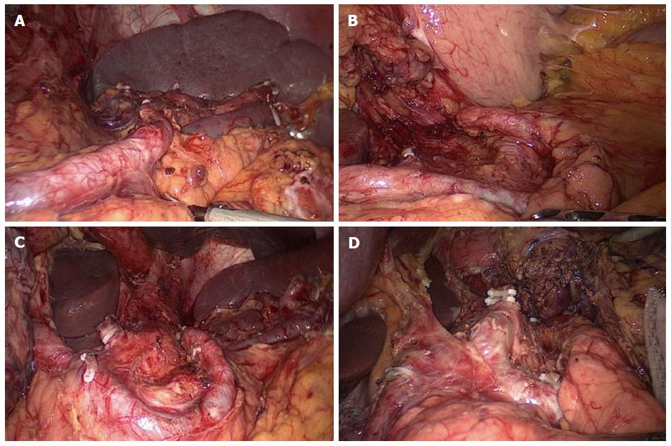
Figure 3 Relation between the course of the splenic artery trunk and the pancreas.
A: Type I; B: Type II; C: Type III; D: Type IV.
- Citation: Zheng CH, Xu M, Huang CM, Li P, Xie JW, Wang JB, Lin JX, Lu J, Chen QY, Cao LL, Lin M. Anatomy and influence of the splenic artery in laparoscopic spleen-preserving splenic lymphadenectomy. World J Gastroenterol 2015; 21(27): 8389-8397
- URL: https://www.wjgnet.com/1007-9327/full/v21/i27/8389.htm
- DOI: https://dx.doi.org/10.3748/wjg.v21.i27.8389


