Copyright
©The Author(s) 2015.
World J Gastroenterol. Jul 21, 2015; 21(27): 8256-8261
Published online Jul 21, 2015. doi: 10.3748/wjg.v21.i27.8256
Published online Jul 21, 2015. doi: 10.3748/wjg.v21.i27.8256
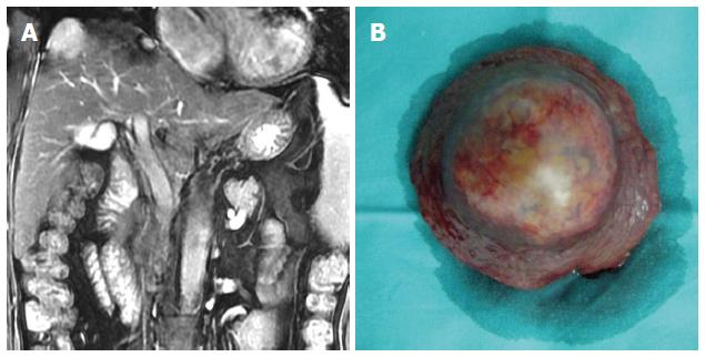
Figure 1 Patient 1.
A 55-year-old woman with a 30-year-history of hepatitis B virus infection presented with persistent right upper-quadrant pain. Her performance status was 2, her AFP was > 1000 μg/L and her liver function was Child-Pugh A. She was diagnosed with a Barcelona Clinic Liver Cancer stage C hepatocellular carcinoma (HCC) with compensated cirrhosis. A: MRI showed a single 3.0-cm tumor in segment 8 with exophytic growth towards the diaphragmatic dome; B: She underwent curative liver resection for HCC in August 2007, and is still alive and disease-free.
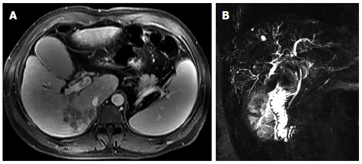
Figure 2 Patient 2.
A 42-year-old man presented with yellowish discoloration of skin. Liver function tests revealed obstructive jaundice. His performance status was 2, his AFP was 512 μg/L and his liver function was Child-Pugh B. He was diagnosed with a Barcelona Clinic Liver Cancer stage C hepatocellular carcinoma with biliary invasion. A: CT scan showed a 5.0-cm tumor with ill-defined margins in segment 6; B: MRCP showed a filling defect in the upper common bile duct.
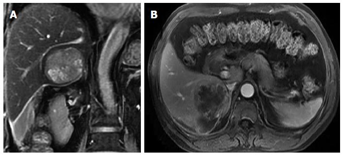
Figure 3 Patient 3.
A 60-year-old man with documented hepatitis B virus infection complained of sudden right abdominal pain and temporary loss of consciousness. His performance status was 3 and his liver function was Child-Pugh A. He was diagnosed with hemorrhagic shock and a Barcelona Clinic Liver Cancer stage D hepatocellular carcinoma (HCC) presenting with spontaneous rupture. A and B: Magnetic resonance imaging showed a single 5.5-cm tumor in segment 6 and homogeneous liquid area around and below the right liver. He underwent emergency liver resection in January 2011 and subsequent abdominal wall metastasis resection in December 2011. He is still alive but he has developed intrahepatic recurrence of HCC.
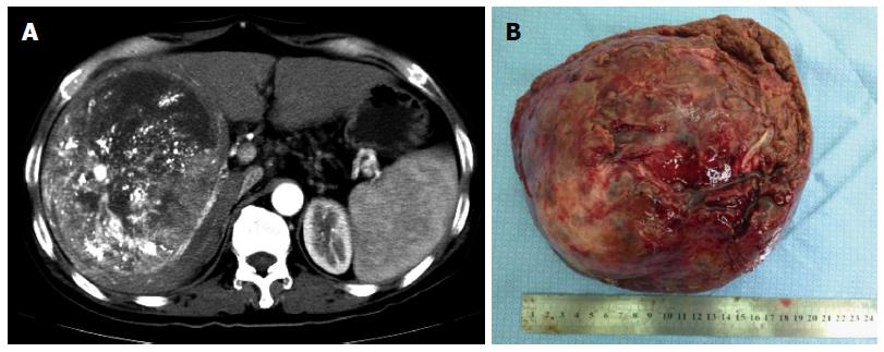
Figure 4 Patient 4.
A 54-year-old man with long-standing chronic hepatitis B virus (HBV) infection and cirrhosis was diagnosed with hepatocellular carcinoma (HCC) during an annual routine check-up. Six weeks before his hospitalization in our center, he received transcatheter arterial chemoembolization for HCC. His performance status was 0 and his liver function was Child-Pugh A. He was diagnosed with a Barcelona Clinic Liver Cancer stage A? B? HCC and HBV-related cirrhosis. A: Computed tomography scan showed a single 17.5-cm tumor in segments 5, 6, 7, and 8 with well-defined margins and sporadic lipiodol depositions within the tumor; H: He underwent curative liver resection of HCC in July 2012, and is still alive.
- Citation: Yang T, Lau WY, Zhang H, Huang B, Lu JH, Wu MC. Grey zone in the Barcelona Clinic Liver Cancer Classification for hepatocellular carcinoma: Surgeons’ perspective. World J Gastroenterol 2015; 21(27): 8256-8261
- URL: https://www.wjgnet.com/1007-9327/full/v21/i27/8256.htm
- DOI: https://dx.doi.org/10.3748/wjg.v21.i27.8256


