Copyright
©The Author(s) 2015.
World J Gastroenterol. Jun 28, 2015; 21(24): 7443-7456
Published online Jun 28, 2015. doi: 10.3748/wjg.v21.i24.7443
Published online Jun 28, 2015. doi: 10.3748/wjg.v21.i24.7443
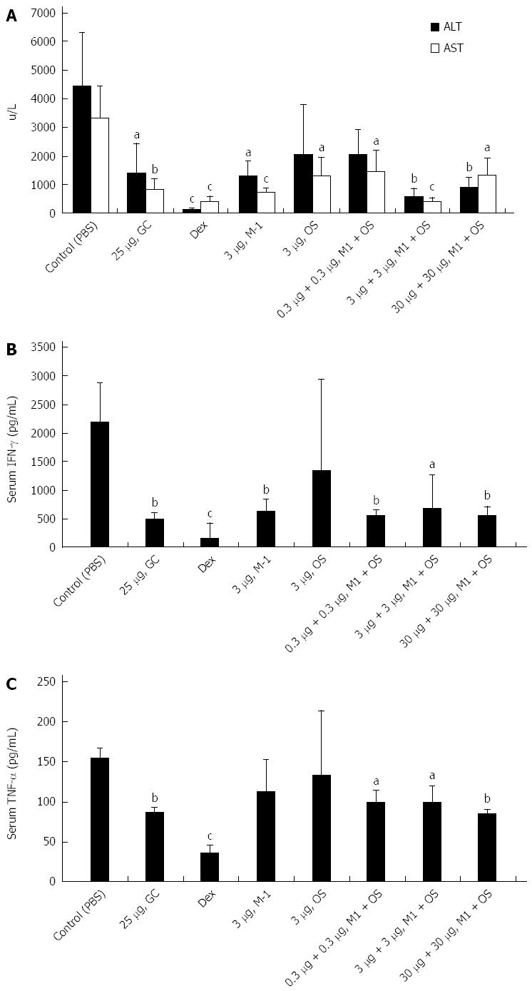
Figure 1 Effect of soy extracts on concanavalin A immune mediated liver injury.
A: Combination of soy extracts in doses of 3 μg and 30 μg of both OS and M1, administered to mice injected with concanavalin A (ConA), significantly decrease aspartate aminotransferase and alanine aminotransferase serum levels (aP < 0.04, bP < 0.01, cP < 0.004 vs control); B: The combination of OS and M1 soy extracts in all doses and 3 μg M1 were associated with significant reduction of serum interferon gamma levels measured by ELISA at the end of the treatment period (aP < 0.02, bP < 0.005, cP = 0.001 vs control); C: All combinations of soy extracts were associated with a significant decrease in serum TNF-α serum levels measured by ELISA at the end of the study (aP < 0.005, bP = 0.0001, cP = 0.00001 vs control). AST: Aspartate aminotransferase; ALT: Alanine aminotransferase.
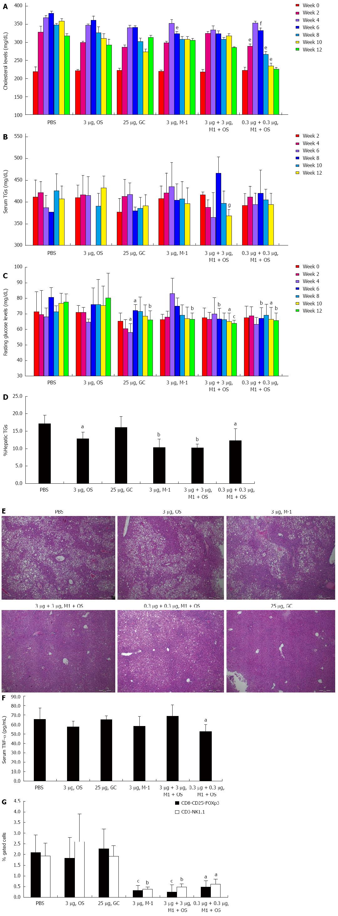
Figure 2 Effect of soy extracts on a high fat diet mouse model of non-alcoholic steatohepatitis.
A: Serum cholesterol levels were measured every two weeks in all groups. The combination of 0.3 μg of OS and M1 was associated with significant a decrease of serum cholesterol level compared to the control group (eP < 0.05, fP < 0.01 vs control); B: Serum triglyceride levels were measured every two weeks in all groups. The combination of 3 μg of each OS and M1 serum levels on week 12 (gP < 0.03 vs control); C: Fasting serum glucose levels were measured every two weeks in all groups. The combination of OS and M1 improved fasting glucose levels as evident from week number 6. A dose of 3 μg of M1 significantly improved fasting glucose level at week 12 (aP < 0.04, bP < 0.008, cP < 0.0005 vs control); D: Hepatic triglyceride content was measured in all mice at the end of the study. The combination of OS and M1, and M1 alone, were associated with significant reduction of hepatic fat content (aP < 0.05, bP < 0.003 vs control); E: Representative H&E (magnification × 10) sections from liver biopsies performed in all groups at the end of the treatment period; F: Serum TNF-α levels were measured by ELISA at the end of the study. A significant reduction was noted in mice treated with the combination of 3 μg M1 and 3 μg OS (aP < 0.05 vs control); G: FACS analysis was performed on splenocytes for NKT and CD8+CD25+Foxp3+ regulatory T lymphocytes. Both the M1 + OS combination and the M1 alone treated group showed a significant reduction in these subsets (aP < 0.002, bP < 0.0009, cP = 0.0003 vs control).
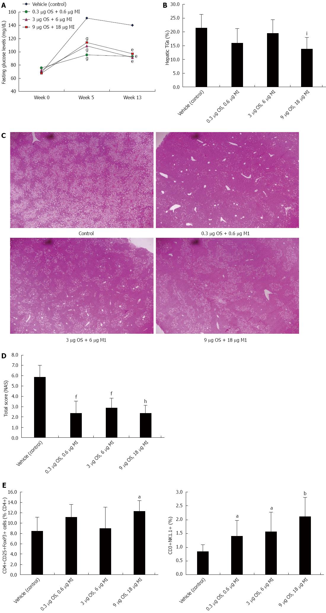
Figure 3 Effect of soy extracts on the high-fat diet/MCD mouse model of non-alcoholic steatohepatitis.
A: The effect of oral administration of soy extracts on fasting serum glucose levels measured on weeks 0, 7 and 13. Glucose levels improved in all treated groups compared to the controls, and the effect was more profound in the 3 μg M1 and 6 μg OS, and 9 μg M1 and 18 μg OS treated groups (eP < 0.01, gP < 0.001 vs control); B: Hepatic triglyceride content was measured in all mice at the end of the study. The combination of 9 μg M1 and 18 μg OS treated groups was associated with significant reduction of hepatic fat content (iP < 0.05 vs control); C: Representative HE (magnification × 10) sections from liver biopsies performed from all groups at the end of the treatment period; D: The pathological NAS score was performed on liver sections from all groups at the end of treatment. A significant improvement was noted in mice in all treated groups (fP < 0.001, hP = 0.0001 vs control); E: FACS analysis was performed on splenocytes for NKT and CD4+CD25+Foxp3+ regulatory T lymphocytes. Both regulatory lymphocyte subsets significantly increased in the spleen of mice treated with the combination of 9 μg M1 and 18 μg OS (aP < 0.05, bP < 0.005 vs control).
- Citation: Khoury T, Ben Ya'acov A, Shabat Y, Zolotarovya L, Snir R, Ilan Y. Altered distribution of regulatory lymphocytes by oral administration of soy-extracts exerts a hepatoprotective effect alleviating immune mediated liver injury, non-alcoholic steatohepatitis and insulin resistance. World J Gastroenterol 2015; 21(24): 7443-7456
- URL: https://www.wjgnet.com/1007-9327/full/v21/i24/7443.htm
- DOI: https://dx.doi.org/10.3748/wjg.v21.i24.7443


