Copyright
©The Author(s) 2015.
World J Gastroenterol. Jun 14, 2015; 21(22): 6974-6981
Published online Jun 14, 2015. doi: 10.3748/wjg.v21.i22.6974
Published online Jun 14, 2015. doi: 10.3748/wjg.v21.i22.6974
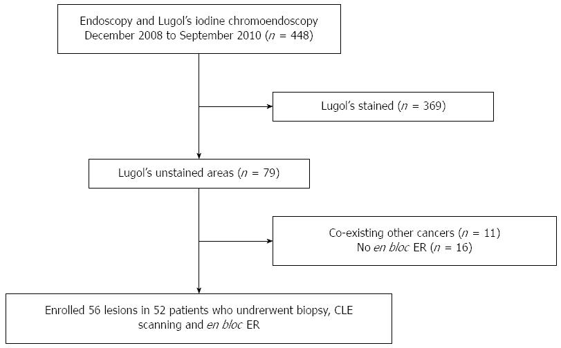
Figure 1 Flow chart of lesion recruitment.
All the endoscopic procedures were performed with the patients under conscious sedation with intravenous midazolam, propofol, fentanyl, or pethidine. Cardiorespiratory function was continually monitored throughout the procedure. CLE: Confocal laser endomicroscopy; ER: Endoscopic resection.
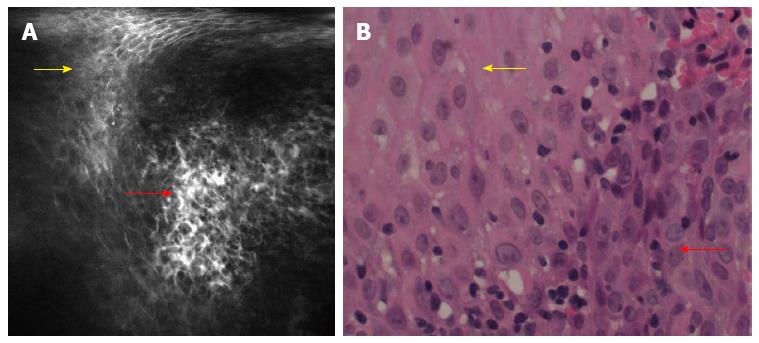
Figure 2 Confocal laser endomicroscopy images of esophageal superficial squamous cell neoplasia.
A: Confocal laser endomicroscopy (CLE) scanning; squamous cells (yellow arrow) are homogeneous, while the indicated squamous cells (red arrow) are irregularly arranged with a distinct size and morphology. Capillary leakage of fluorescein sodium is observed; B: Pathological images; the yellow arrow indicates homogeneous cells; the red arrow indicates disordered cell arrangement. The cells have a distinct size and morphology, which is in accordance with CLE.
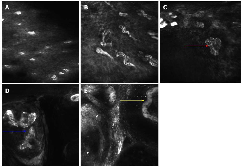
Figure 3 Confocal laser endomicroscopy images showing different intraepithelial papillary capillary loop changes in the esophageal lesions.
A: Regular squamous esophageal epithelium with regular intraepithelial papillary capillary loops (IPCLs) and epithelial cells; B: Some tortuous IPCLs are seen in the non-neoplastic inflammatory lesion; C: Various shapes of twisted IPCLs (red arrow) are seen in the low-grade intraepithelial neoplasia lesion; D: Obvious caliber and shape changes with a larger diameter IPCL (blue arrow) are seen in the high-grade intraepithelial neoplasia lesion; E: Tumor vessels (yellow arrow) are seen in esophageal squamous cell carcinoma.
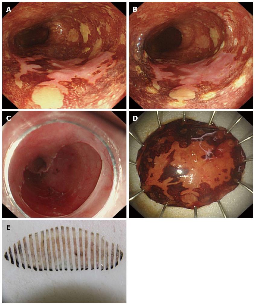
Figure 4 En bloc endoscopic resection for esophageal lesions.
A: Lugol’s iodine chromoendoscopy showed an unstained lesion located in the esophagus; B: The marks surrounding the lesion are at least 5 mm away from the lesion; C: Upper gastrointestinal endoscopy showed an artificial ulceration after ESD; D: En bloc resected specimen. 3.0 cm × 3.0 cm; E: The resected specimen was fixed in 10% formaldehyde solution and continuously sliced into 2 mm sections from the proximal end to the distal end for pathology.
- Citation: Huang J, Yang YS, Lu ZS, Wang SF, Yang J, Yuan J. Detection of superficial esophageal squamous cell neoplasia by chromoendoscopy-guided confocal laser endomicroscopy. World J Gastroenterol 2015; 21(22): 6974-6981
- URL: https://www.wjgnet.com/1007-9327/full/v21/i22/6974.htm
- DOI: https://dx.doi.org/10.3748/wjg.v21.i22.6974


