Copyright
©The Author(s) 2015.
World J Gastroenterol. Jun 7, 2015; 21(21): 6561-6571
Published online Jun 7, 2015. doi: 10.3748/wjg.v21.i21.6561
Published online Jun 7, 2015. doi: 10.3748/wjg.v21.i21.6561
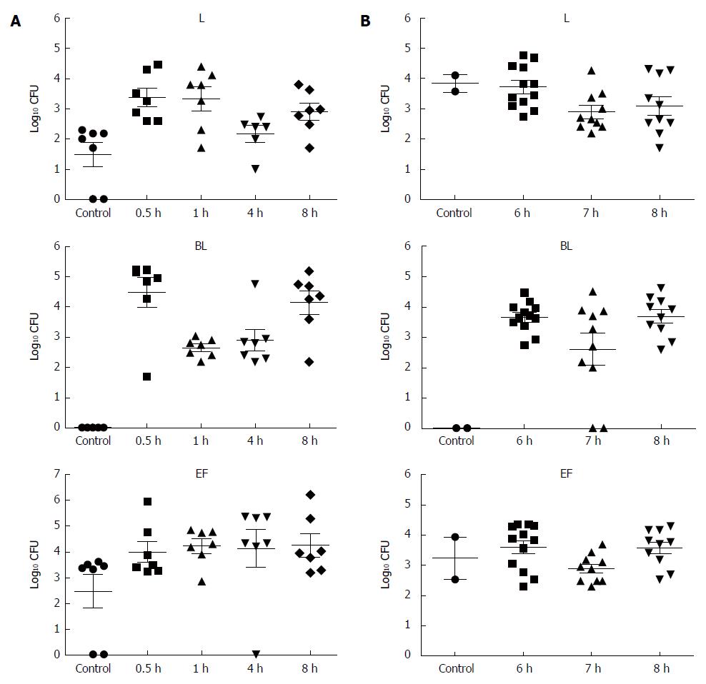
Figure 1 Colonization of BIFICO strain in mouse stomach.
A: Stomach loads of Lactobacilli acidophilus (L), Bifidobacteria longum (BL) and Enterococci faecali (EF) at the indicated times after mice were administrated with a single dose of BIFICO (L, 107 CFU; BL, 107 CFU; EF, 107 CFU); B: Stomach loads of L, BL and EF at the indicated times after mice were administrated with BIFICO (L, 107 CFU; BL, 107 CFU; EF, 107 CFU) once a day for 5 d.
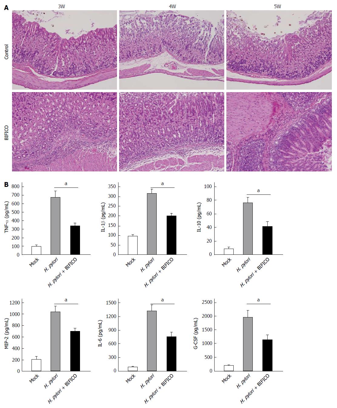
Figure 2 BIFICO ameliorates Helicobacter pylori induced gastritis in mice.
A: Stomach histopathology of mice with Helicobacter pylori (H. pylori) infection was analyzed with hematoxylinand eosin (HE) staining at the indicated times after treatment with BIFICO; B: Production of cytokines and chemokines including TNF-α, IL-1β, IL-10, IL-6, G-CSF and MIP-2 in mouse stomach stimulated with H. pylori. The levels of cytokines and chemokines in the extracts of homogenized tissues were measured with the MILLIPLEX Mouse Cytokine and Chemokine Magnetic Bead Panel. Data shown are representative of three independent experiments; aP < 0.05, H. pylori vs H. pylori + BIFICO (t test).
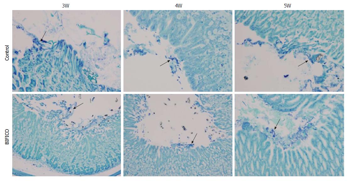
Figure 3 BIFICO does not suppress Helicobacter pylori colonization in mouse stomach.
Stomach histopathology of mice with Helicobacter pylori infection was analyzed with Giemsa staining at the indicated times after treatment with BIFICO.
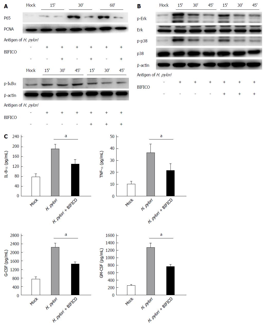
Figure 4 Nuclear factor-κB and MAPK signaling activation in human gastric epithelial cell GES-1 induced by Helicobacter pylori is suppressed by BIFICO.
A, B: Epithelial GES-1 cells treated with or without BIFICO were stimulated by inactivated Helicobacter pylori (H. pylori) (MOI = 5) for the indicated times. The cell lysates (A) and nuclear extracts (B) were analyzed by immunoblotting with the indicated antibodies. A set of representative results from three independent experiments was presented; C: ELISA results for IL-8, TNF-α, G-CSF, and GM-CSF in supernatants of GES-1 treated with or without BIFICO, which were stimulated with inactivated H. pylori for 24 h. Data shown are representative of three independent experiments. SD is indicated; aP < 0.05, H. pylori vs H. pylori + BIFICO (t test).
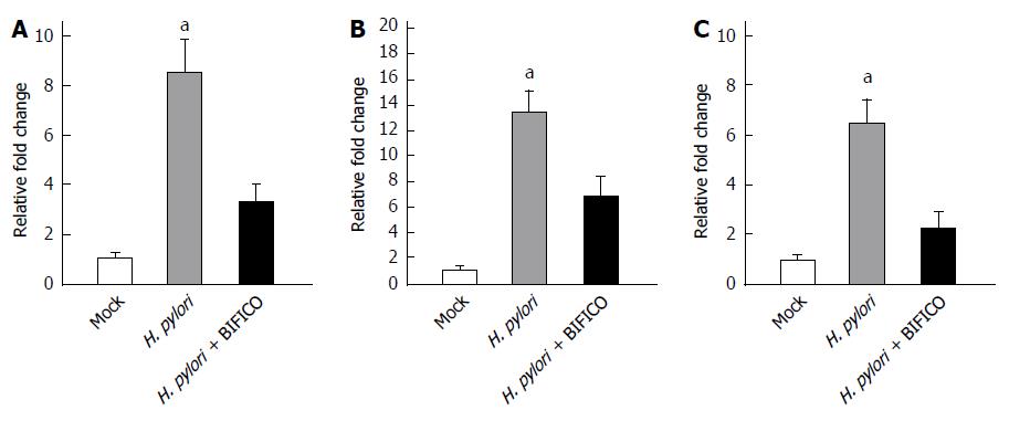
Figure 5 TLR2, TLR4 and Myd88 overexpression in mouse stomach induced by Helicobacter pylori is suppressed by BIFICO.
A-C: Quantitative reverse transcription-polymerase chain reaction analysis of TLR2 (A), TLR4 (B) and Myd88 (C) mRNAs in mouse stomach stimulated with Helicobacter pylori (H. pylori). aP < 0.05, H. pylori vs H. pylori + BIFICO (t test).
-
Citation: Yu HJ, Liu W, Chang Z, Shen H, He LJ, Wang SS, Liu L, Jiang YY, Xu GT, An MM, Zhang JD. Probiotic BIFICO cocktail ameliorates Hel
icobacter pylori induced gastritis. World J Gastroenterol 2015; 21(21): 6561-6571 - URL: https://www.wjgnet.com/1007-9327/full/v21/i21/6561.htm
- DOI: https://dx.doi.org/10.3748/wjg.v21.i21.6561


