Copyright
©The Author(s) 2015.
World J Gastroenterol. Jun 7, 2015; 21(21): 6550-6560
Published online Jun 7, 2015. doi: 10.3748/wjg.v21.i21.6550
Published online Jun 7, 2015. doi: 10.3748/wjg.v21.i21.6550
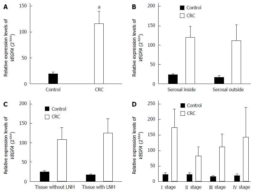
Figure 1 Expression levels of VEGFA mRNA assessed by quantitative reverse transcription-polymerase chain reaction.
A: Colorectal cancer (CRC) tissues and normal intestinal mucosa tissues; B: Tissues inside and outside the serosal layer; C: Tissues with and without lymph node metastasis (LNM); D: Tissues of tumor-node-metastasis (TNM) stages. aP < 0.05 vs control.
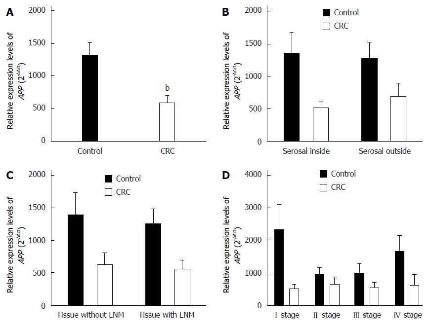
Figure 2 Expression levels of APP mRNA assessed by quantitative reverse transcription-polymerase chain reaction.
A: Colorectal cancer (CRC) tissues and normal intestinal mucosa tissues; B: Tissues inside and outside the serosal layer; C: Tissues with and without lymph node metastasis (LNM); D: Tissues of tumor-node-metastasis (TNM) stages. bP < 0.01 vs control.
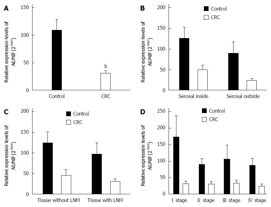
Figure 3 Expression levels of NUMB mRNA assessed by quantitative reverse transcription-polymerase chain reaction.
A: Colorectal cancer (CRC) tissues and normal intestinal mucosa tissues; B: Tissues inside and outside the serosal layer; C: Tissues with and without lymph node metastasis (LNM); D: Tissues of tumor-node-metastasis (TNM) stages. bP < 0.01 vs control.
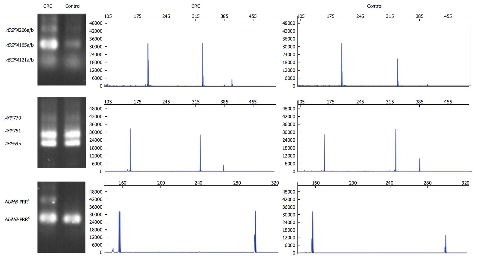
Figure 4 Electrophoregram of alternative splicing variants in VEGFA, APP and NUMB.
CRC: Colorectal cancer.
Figure 5 Expression levels of alternative splice variants using polymerase chain reaction-restriction fragment length polymorphism analysis.
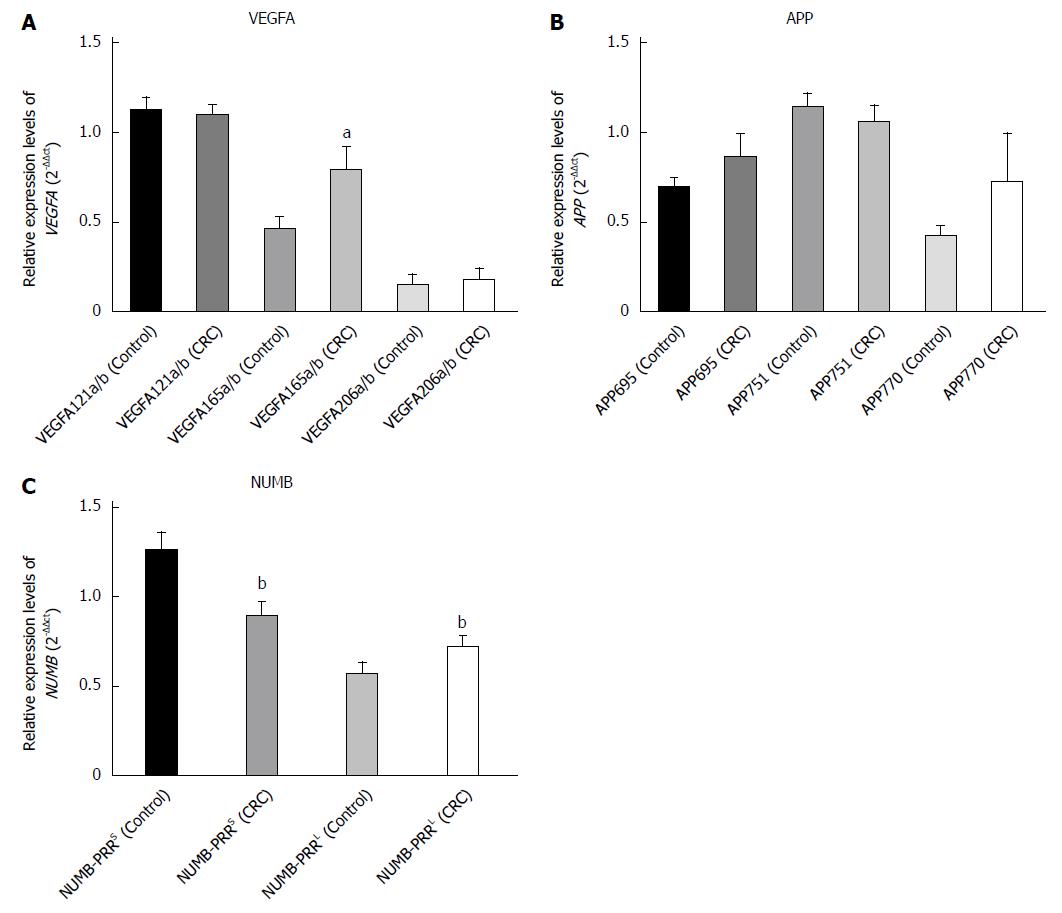
Expression levels of A: VEGFA; B: APP; and C: NUMB alternative splice variants in colorectal cancer (CRC) tissues and normal intestinal mucosa tissues. aP < 0.05 and bP < 0.01 vs the control group.
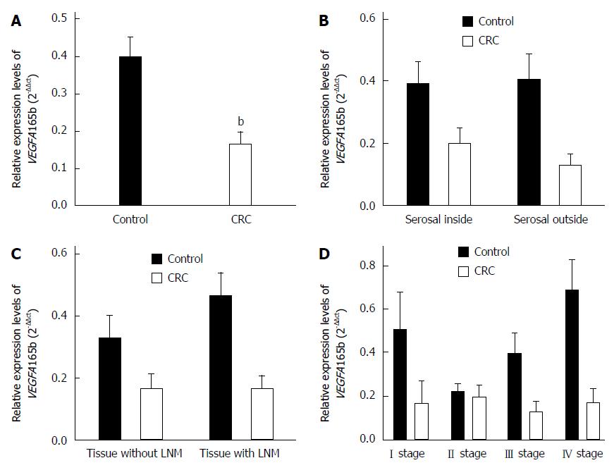
Figure 6 Expression levels of VEGFA165b assessed by quantitative reverse transcription-polymerase chain reaction.
Expression levels of VEGFA165b in A: Colorectal cancer (CRC) tissues and normal intestinal mucosa tissues; B: Tissues inside and outside the serosal layer; C: Tissues with and without lymph node metastasis (LNM); and D: Tissues of tumor-node-metastasis (TNM) stages. bP < 0.01 vs control.
-
Citation: Zhao YJ, Han HZ, Liang Y, Shi CZ, Zhu QC, Yang J. Alternative splicing of
VEGFA ,APP andNUMB genes in colorectal cancer. World J Gastroenterol 2015; 21(21): 6550-6560 - URL: https://www.wjgnet.com/1007-9327/full/v21/i21/6550.htm
- DOI: https://dx.doi.org/10.3748/wjg.v21.i21.6550


