Copyright
©The Author(s) 2015.
World J Gastroenterol. Jun 7, 2015; 21(21): 6526-6542
Published online Jun 7, 2015. doi: 10.3748/wjg.v21.i21.6526
Published online Jun 7, 2015. doi: 10.3748/wjg.v21.i21.6526
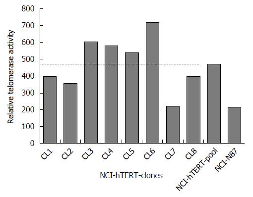
Figure 1 Relative telomerase activity measured for 8 NCI-N87 derived clones (NCI-hTERT-CL1 to NCI-hTERT-CL8), a pool of the remaining hTERT-expressing NCI-N87 derived clones (NCI-hTERT-pool cell line) and for the parental cell line (NCI-N87, ATCC CRL-5822).
These values were obtained using the TeloTAGGG Telomerase PCR ELISAPLUS (Roche Diagnostics), according to the manufacturer’s instructions. The dashed line indicates the average level of the relative telomerase activity measured for the clones.
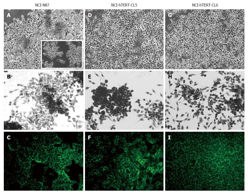
Figure 2 Microscopy analysis of NCI-N87 and NCI-hTERT-clones CL5 and CL6 cell lines.
Confluent cultures (A, D, G); square in (A and B), subconfluent cultures (E and H); E-cadherin immunodetection (C, F and I) (green; 1:1000, E-cadherin mAb plus FITC-conjugated secondary Ab). White bar: 100 μm.
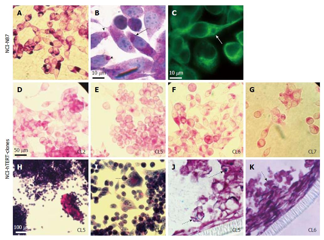
Figure 3 Cell staining analysis for mucins detection on the NCI-N87 parental cell line (A-C) and NCI-hTERT-CL2 (D), CL5 (E, H, I and J), CL6 (F and K) and -CL7 (G).
For neutral mucins detection (stained in pink): PAS-staining (A, D-I); and PAS/haematoxylin staining (B). For acidic mucins detection (J and K) PAS/Alcian blue staining (Alcian positive/PAS negative mucins stained in blue; PAS/Alcian positive mucins stained in purple). α-Tubulin immunodetection (C; green) (1:1000 α-Tubulin mAb plus FITC-conjugated secondary Ab). Black arrow, mucus secreting vesicles that are formed in the endoplasmic reticulum vicinity. Arrow heads, mucus secreting vesicles migrating towards the cytoplasmic membrane and being exocytosed. White arrow, mucus secreting vesicles in close interaction with the microtule network. Dashed arrow suggestive of a more differentiated state for the cells. Dotted arrows, acidic mucins staining.
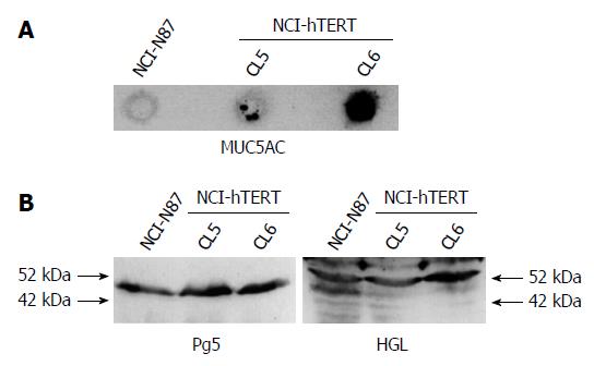
Figure 4 Evaluation of the expression of epithelial gastric markers by the parental NCI-N87 and NCI-hTERT-clones 5 and 6 cell lines.
A: Slot-blot of 100 μg of total protein extracts for detection of mucin 5AC (MUC5AC) with the anti-MUC5AC mAb (1:1000 diluted); B: Western blot of 100 μg of total protein extracts separated in a 12.5% (v/v) SDS-PAGE, with the anti-Pg5 mAb (1:800 diluted) and the anti-HGL pAb (1:800 diluted).
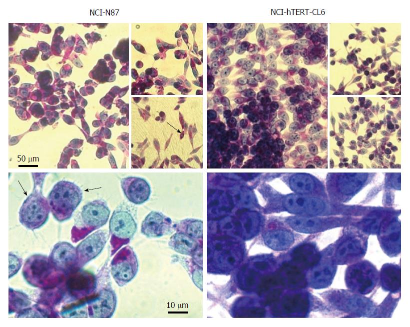
Figure 5 PAS/haematoxylin cell staining analysis for simultaneous detection of mucins (stained in pink) and zymogens (stained in blue) on the NCI-N87 parental cell and the NCI-hTERT-CL6.
The duple PAS/haematoxylin reactivity was observed in only some cell subpopulations of the parental cell line (black arrows) and, in contrast, in all cells of the NCI-hTERT-CL6 cell line.
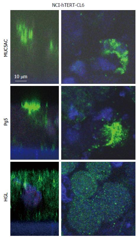
Figure 6 Gastric markers expression by the NCI-hTERT-CL 6 cell line.
Mucin 5AC (MUC5AC), Pg5 and HGL were respectively immunodetected (green) with the anti-MUC5AC mAb (1:25), the anti-Pg5 mAb (1:25) and the anti-HGL pAb (1:25) plus a FITC-conjugated secondary Ab. Nuclei were counterstained with DAPI. Fluorescent signals were recorded by confocal microscopy. Left panels, vertical sections with apical membrane on top of the image. Right panels, horizontal section of same cell sample.
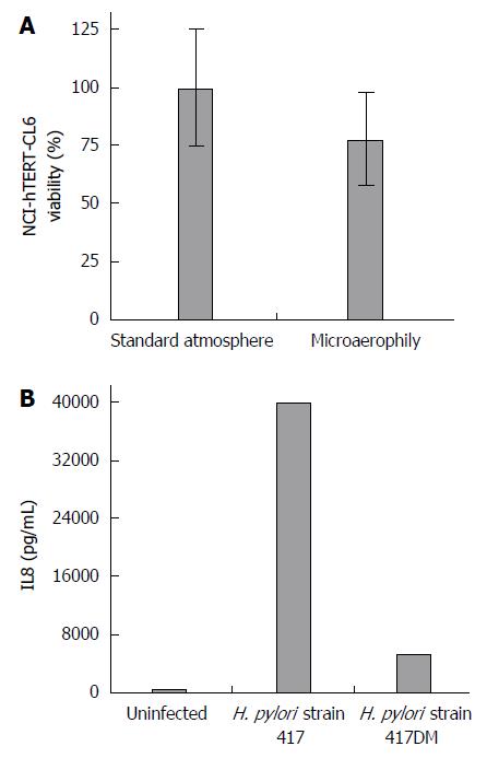
Figure 7 Usefulness of the NCI-hTERT-CL6 cell line for in vitro infection assays with Helicobacter pylori.
A: Viability in percentage of the cells when grown in a microaerobic environment (bacterial growth conditions) for 48 h compared to those grown at standard atmosphere (taken as 100%). Bars and respective error bars are mean ± SD of the values at each condition for twelve observations; B: IL8 secretion by NCI-hTERT-CL6 cells upon infection with the Helicobacter pylori (H. pylori) strain 417 (virulent strain) or the respective double-mutant for homB gene strain (strain 417DM) (less virulent strain), compared to uninfected cells. Bars are values obtained from one observation.
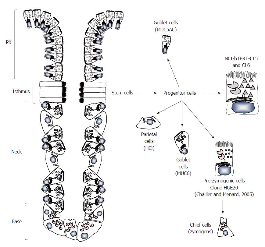
Figure 8 Oxyntic gastric gland unit.
These are organized in four distinctive zones: the pit zone, which is lined by mucin (MUC) 5AC-secreting pit cells; the isthmus, which houses the stem and progenitor cells; the neck zone, which contains MUC6-secreting neck cells and HCl-secreting parietal cells; and the base zone, with the enzyme-secreting chief cells. Gastric epithelial cells turnover is ensured by differentiation bidirectional migration of progenitor cells of the isthmus to the correct place for the emerging adult cell. The new NCI-hTERT-CL5 and CL6 cell lines present dual features of simultaneous production and secretion of MUC5AC (as pit cells) and HGL and Pg5 zymogens (as chief cells), a progenitor-like phenotype.
- Citation: Saraiva-Pava K, Navabi N, Skoog EC, Lindén SK, Oleastro M, Roxo-Rosa M. New NCI-N87-derived human gastric epithelial line after human telomerase catalytic subunit over-expression. World J Gastroenterol 2015; 21(21): 6526-6542
- URL: https://www.wjgnet.com/1007-9327/full/v21/i21/6526.htm
- DOI: https://dx.doi.org/10.3748/wjg.v21.i21.6526


