Copyright
©The Author(s) 2015.
World J Gastroenterol. May 21, 2015; 21(19): 6077-6081
Published online May 21, 2015. doi: 10.3748/wjg.v21.i19.6077
Published online May 21, 2015. doi: 10.3748/wjg.v21.i19.6077
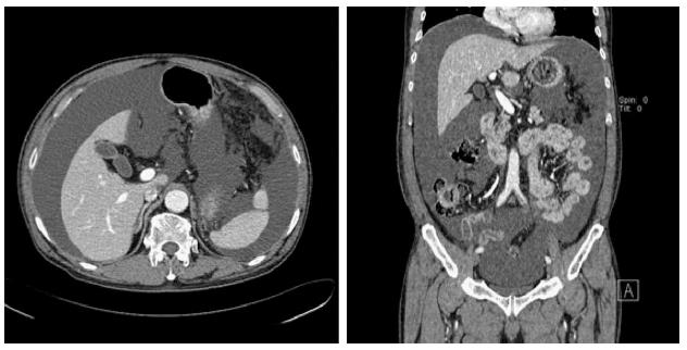
Figure 1 Abdominal computed tomography images showed a large volume of ascites.
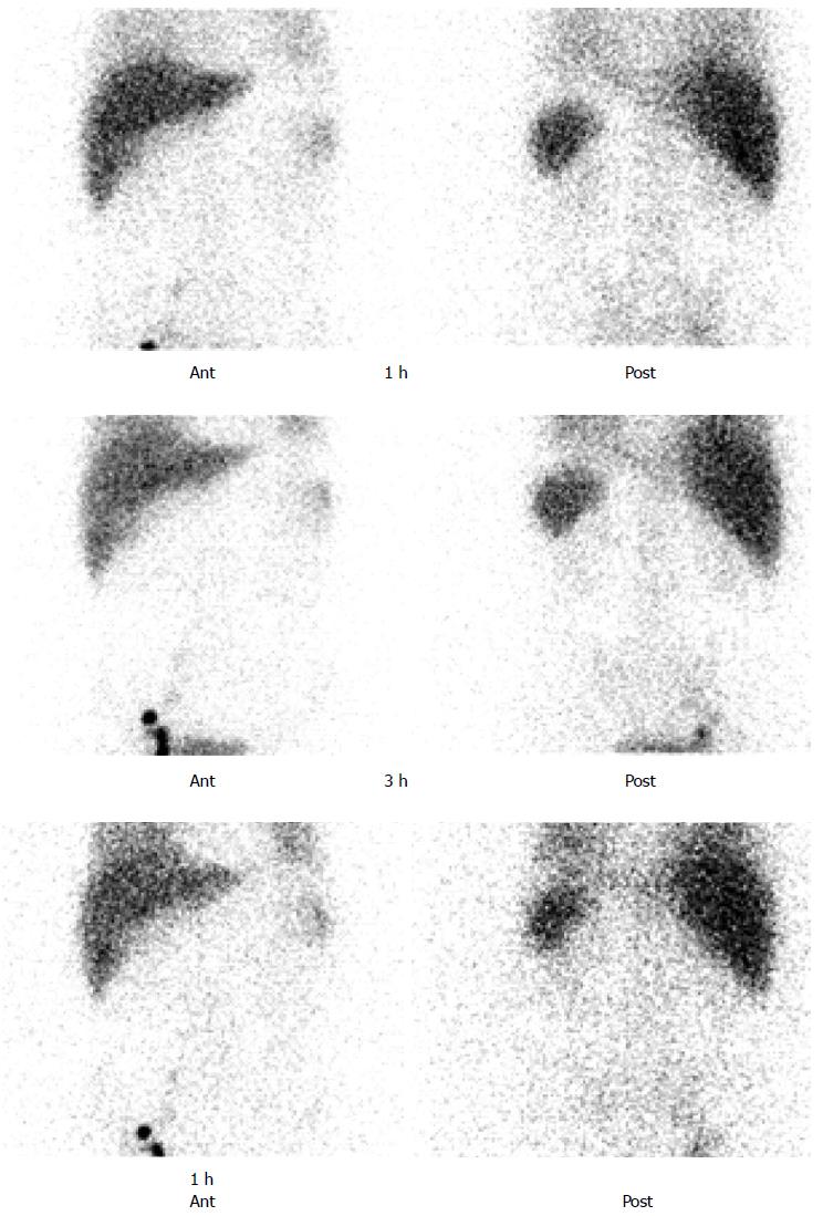
Figure 2 Preoperative lymphoscintigraphy showed no evidence of lymphatic leakage in abdominal cavity.
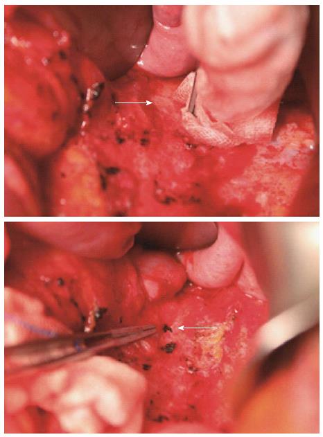
Figure 3 Fistula was found to stem from a 1 mm hole on the left side of the ligated inferior mesenteric artery, and the tract was sutured (white arrows).
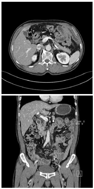
Figure 4 Postoperative abdominal computed tomography images showed no evidence of ascites.
- Citation: Ha GW, Lee MR. Surgical repair of intractable chylous ascites following laparoscopic anterior resection. World J Gastroenterol 2015; 21(19): 6077-6081
- URL: https://www.wjgnet.com/1007-9327/full/v21/i19/6077.htm
- DOI: https://dx.doi.org/10.3748/wjg.v21.i19.6077


