Copyright
©The Author(s) 2015.
World J Gastroenterol. May 21, 2015; 21(19): 6072-6076
Published online May 21, 2015. doi: 10.3748/wjg.v21.i19.6072
Published online May 21, 2015. doi: 10.3748/wjg.v21.i19.6072
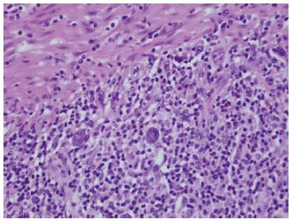
Figure 1 Hematoxylin and eosin staining of the colectomy specimen (magnification × 40).
Scattered Hodgkin/Reed-Sternberg-like cells are present in a polymorphous background of lymphocytes, histiocytes and eosinophils. Muscularis propria is seen in the upper left of the field.
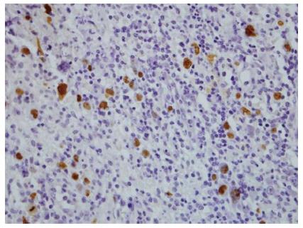
Figure 2 Epstein Barr virus-encoded small RNAs in situ hybridisation of the colectomy specimen (magnification × 40).
The Hodgkin/Reed-Sternberg-like cells show strong nuclear staining, indicating Epstein-Barr virus positivity.
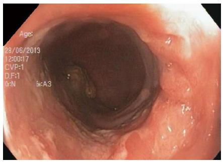
Figure 3 Persistence of ulceration in the sigmoid colon 18 mo post methotrexate and infliximab cessation.
- Citation: Moran NR, Webster B, Lee KM, Trotman J, Kwan YL, Napoli J, Leong RW. Epstein Barr virus-positive mucocutaneous ulcer of the colon associated Hodgkin lymphoma in Crohn’s disease. World J Gastroenterol 2015; 21(19): 6072-6076
- URL: https://www.wjgnet.com/1007-9327/full/v21/i19/6072.htm
- DOI: https://dx.doi.org/10.3748/wjg.v21.i19.6072


