Copyright
©The Author(s) 2015.
World J Gastroenterol. May 21, 2015; 21(19): 5950-5960
Published online May 21, 2015. doi: 10.3748/wjg.v21.i19.5950
Published online May 21, 2015. doi: 10.3748/wjg.v21.i19.5950
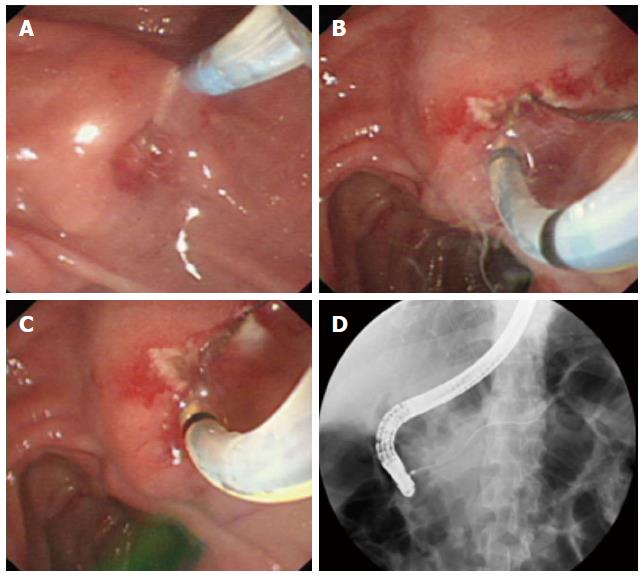
Figure 1 “Needle-knife papillotomy with introduction of a guidewire and pull-sphincterotome” procedure.
A: A small incision was made with the needle-knife (precut papillotomy); B: The needle-knife was exchanged for a pull-sphincterotome with a guidewire, which was carefully advanced into the duct of Santorini through the sphincterotome; C: The sphincterotome advanced along the guidewire; D: Guidewire located in the duct of Santorini.
Figure 2 “Needle-knife papillotomy with introduction of a guidewire, pull-sphincterotome, and guidewire” procedure.
A: A small incision was made with the needle-knife (precut papillotomy); B: The needle-knife was exchanged for a pull-sphincterotome, which was used to enlarge the incision; C: A guidewire was carefully advanced into the duct of Santorini through the sphincterotome; D: Guidewire located in the duct of Santorini.
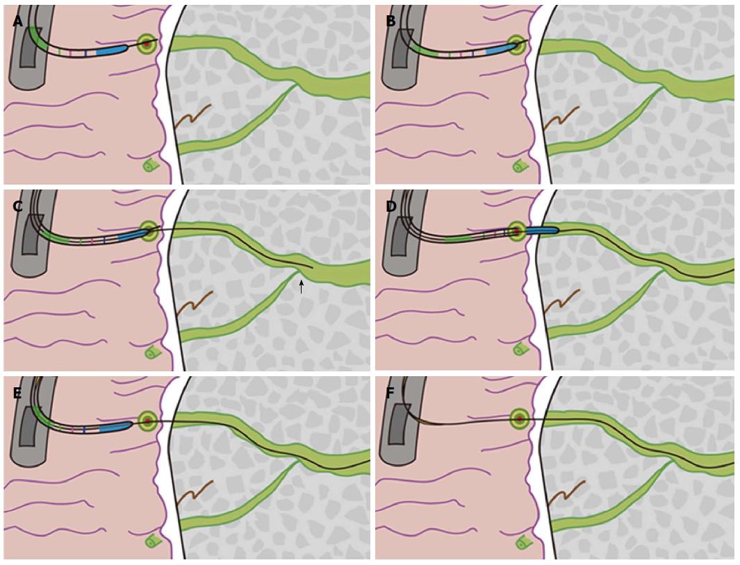
Figure 3 Schematic for the “needle-knife introduction of a guidewire” procedure.
A: The needle tip was inserted into the orifice; B: The needle-knife cannula was placed on the minor papilla orifice; C: The guidewire was carefully advanced through the cannula until it passed the cross-point (black arrow) of the ducts of Wirsung and Santorini; D: The cannula was inserted into the capitular head of the duct of Santorini along the guidewire and advanced; E: The needle-knife was removed; F: The guidewire was left in place.
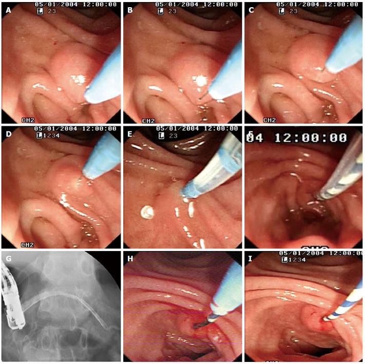
Figure 4 “Needle-knife introduction of a guidewire” procedure.
A: The cannula of the needle-knife approaching the minor papilla orifice; B: The needle tip was extended 3 to 5 mm beyond the cannula tip and aimed at the orifice; C, D: The needle tip was inserted into the orifice; E: The cannula of the needle-knife was placed on the minor papilla orifice and a guidewire was advanced through the cannula; F: The cannula was inserted into the capitular head of the duct of Santorini along the guidewire and advanced continually; G: Contrast material was injected and the location within the duct of Santorini was confirmed; H: The needle-knife was removed; I: The guidewire was left in place.
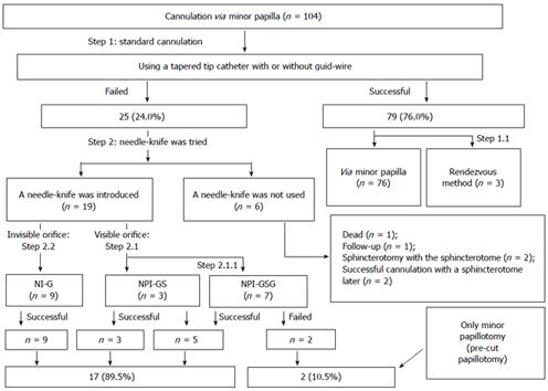
Figure 5 Cannulation procedures via the minor papilla.
NI-G: Needle-knife introduction of a guidewire; NPI-GS: Needle-knife papillotomy with introduction of a guidewire and pull-sphincterotome; NPI-GSG: Needle-knife papillotomy with introduction of a guidewire, pull-sphincterotome, and guidewire.
-
Citation: Wang W, Gong B, Jiang WS, Liu L, Bielike K, Xv B, Wu YL. Endoscopic treatment for pancreatic diseases: Needle-knife-guided cannulation
via the minor papilla. World J Gastroenterol 2015; 21(19): 5950-5960 - URL: https://www.wjgnet.com/1007-9327/full/v21/i19/5950.htm
- DOI: https://dx.doi.org/10.3748/wjg.v21.i19.5950


