Copyright
©The Author(s) 2015.
World J Gastroenterol. May 14, 2015; 21(18): 5635-5640
Published online May 14, 2015. doi: 10.3748/wjg.v21.i18.5635
Published online May 14, 2015. doi: 10.3748/wjg.v21.i18.5635
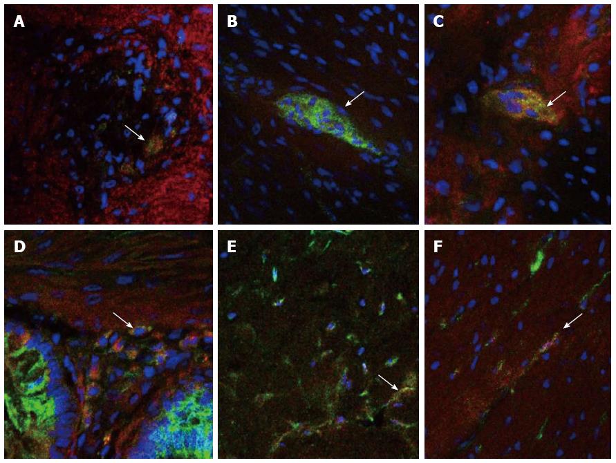
Figure 1 Immunofluorescent staining in normal control tissue of HCN2 channels (green), co-localised (arrows) with neuronal marker HuC/D (red) (A) and c-kit (red) (D), HCN3 channels (red) co-localised with PGP9.
5 (green) (B), and TMEM (green) (E), and HCN4 channels (green) co-localised with HuC/D (red) (C) and c-kit (red) (F). Nuclei were stained with DAPI (blue).
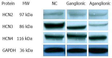
Figure 2 Western blot of HCN2, HCN3 and HCN4 protein expression.
HCN2 protein expression was low in normal controls and similarly expressed in ganglionic and aganglionic specimens. HCN3 protein was highly expressed in normal controls, decreased in ganglionic specimens with a further decrease evident in aganglionic segments. HCN4 protein was highly expressed in normal controls and similarly expressed in both ganglionic and aganglionic specimens. The loading control GAPDH was similarly expressed in normal controls, ganglionic and aganglionic specimens.
- Citation: O’Donnell AM, Coyle D, Puri P. Decreased expression of hyperpolarisation-activated cyclic nucleotide-gated channel 3 in Hirschsprung’s disease. World J Gastroenterol 2015; 21(18): 5635-5640
- URL: https://www.wjgnet.com/1007-9327/full/v21/i18/5635.htm
- DOI: https://dx.doi.org/10.3748/wjg.v21.i18.5635


