Copyright
©The Author(s) 2015.
World J Gastroenterol. Apr 14, 2015; 21(14): 4419-4422
Published online Apr 14, 2015. doi: 10.3748/wjg.v21.i14.4419
Published online Apr 14, 2015. doi: 10.3748/wjg.v21.i14.4419
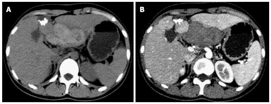
Figure 1 Radiographic findings.
A: A non-enhanced computed tomography (CT) scan revealed two masses in the liver, one of which contained a high-density nodule; B: A contrast-enhanced CT scan obtained during the hepatic arterial phase showed no enhancement in or around the masses.
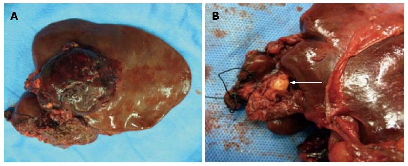
Figure 2 Intraoperative findings.
A: A solid mass localized in segment III; B: A cystic mass with a hard nodule (arrow) localized in segment IV.
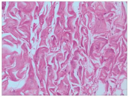
Figure 3 Histologic findings.
A histologic section from the nodule revealed thickened, banded and sheet-like collagenous fibrous tissue (Hematoxylin and eosin stain, magnification × 200).
- Citation: Zheng ZJ, Zhang S, Cao Y, Pu GC, Liu H. Collagenous nodule mixed simple cyst and hemangioma coexistence in the liver. World J Gastroenterol 2015; 21(14): 4419-4422
- URL: https://www.wjgnet.com/1007-9327/full/v21/i14/4419.htm
- DOI: https://dx.doi.org/10.3748/wjg.v21.i14.4419


