Copyright
©The Author(s) 2015.
World J Gastroenterol. Apr 14, 2015; 21(14): 4373-4378
Published online Apr 14, 2015. doi: 10.3748/wjg.v21.i14.4373
Published online Apr 14, 2015. doi: 10.3748/wjg.v21.i14.4373
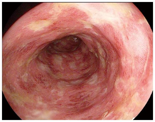
Figure 1 Erythematous mucosa, loss of normal vascular pattern, multiple ulcers most consistent with IpiColitis.
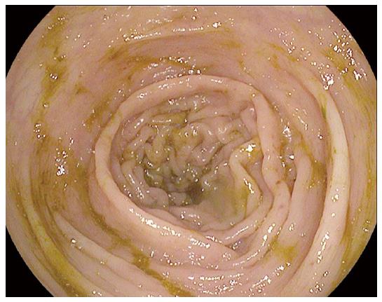
Figure 2 Normal colonoscopy.
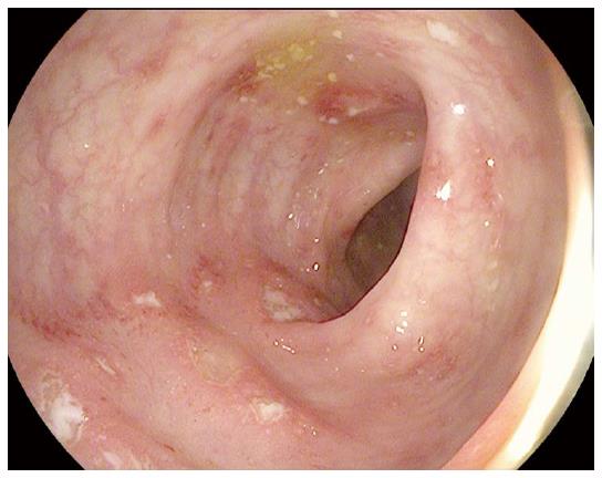
Figure 3 Erythema with ulceration and loss of vascular pattern.
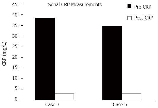
Figure 4 Graph demonstrating normalization of C-reactive protein levels after treatment in two of the seven cases.
CRP: C-reactive protein.
- Citation: Rastogi P, Sultan M, Charabaty AJ, Atkins MB, Mattar MC. Ipilimumab associated colitis: An IpiColitis case series at MedStar Georgetown University Hospital. World J Gastroenterol 2015; 21(14): 4373-4378
- URL: https://www.wjgnet.com/1007-9327/full/v21/i14/4373.htm
- DOI: https://dx.doi.org/10.3748/wjg.v21.i14.4373


