Copyright
©The Author(s) 2015.
World J Gastroenterol. Mar 21, 2015; 21(11): 3394-3401
Published online Mar 21, 2015. doi: 10.3748/wjg.v21.i11.3394
Published online Mar 21, 2015. doi: 10.3748/wjg.v21.i11.3394
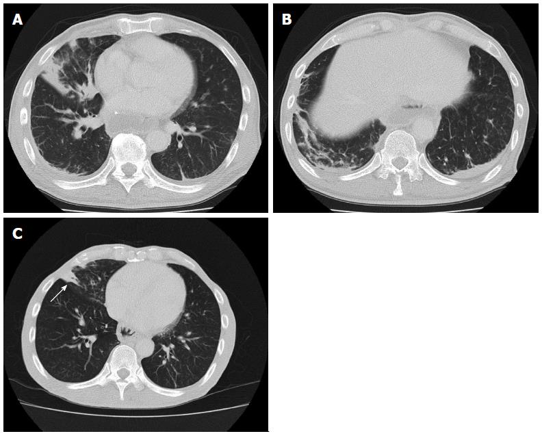
Figure 1 Unenhanced computed tomography after esophagectomy.
A: Computed tomography (CT) revealed consolidation in the right middle lobe; B: CT simultaneously demonstrated consolidation in the right lower lobe (A and B); C: Although there was no evidence of newly developed pneumonia, consolidation remained in the right middle lobe 14 mo after esophagectomy (arrow).
Figure 2 Imaging findings before bronchial arterial embolization.
A: Bronchoscopy revealed hemosputum in the entrance of the right middle lobe bronchus (yellow arrow) extending down to the lower lobe bronchus (green arrow); B, C: Computed tomography and chest radiography revealed a massive infiltrative shadow in the right middle lobe with atelectasis in the right lower lobe.
Figure 3 Computed tomography arteriography.
A: Computed tomography (CT) arteriography revealed a tortuous artery ascending from the right inferior phrenic artery that arose directly from the aorta (arrows); B: CT arteriography demonstrated several tortuous arteries arising from the left subclavian artery that descended obliquely along the right main bronchus (arrows); C: A collateral right bronchial artery (BA) arising from the inferior phrenic artery. Right inferior phrenic arteriography showed an abnormal tortuous artery (arrows); D: Ectopic BAs arising from the left subclavian artery. Left subclavian arteriography showed abnormal tortuous arteries arising from a root of the left subclavian artery (arrows).
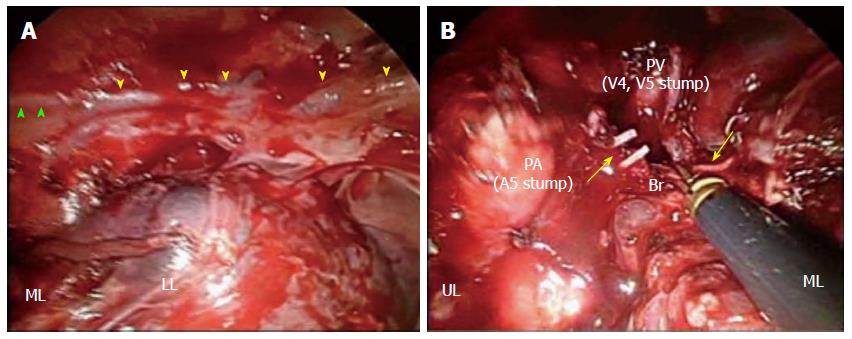
Figure 4 Intraoperative findings.
A: The presence of a collateral bronchial artery (BA) arising from the right inferior phrenic artery running along the phrenic nerve (green arrowheads) was confirmed; the tissue was black in color due to the first bronchial arterial embolization procedure (yellow arrowheads). B: Engorged right BAs streaming into the right middle lobe were dissected at the level of bifurcation of the middle lobe bronchus (arrows). UL: Upper lobe; ML: Middle lobe; LL: Lower lobe; Br: Middle lobe bronchus; PA: Pulmonary artery; PV: Pulmonary vein.
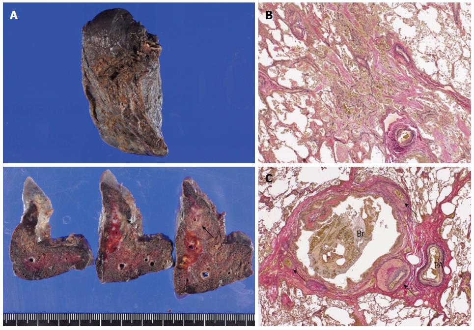
Figure 5 Gross and histopathological findings of the resected specimen.
A: An ill-demarcated whitish area with a brownish tint was noted on the cut surface of the resected lung (arrows). Macroscopically, the bleeding origin was not detected; B: Organizing pneumonia with the accumulation of hemosiderin-laden macrophages was observed [Elastica van Gieson stain (EVG), magnification × 24]; C: The bronchus was enlarged, and engorged bronchial artery with medial hypertrophy and overgrowth of small branches were identified near the bronchus (arrows) (EVG, magnification × 24). Br: Bronchus; PA: Pulmonary artery.
- Citation: Kitajima T, Momose K, Lee S, Haruta S, Ueno M, Shinohara H, Fujimori S, Fujii T, Takei R, Kohno T, Udagawa H. Bronchial bleeding caused by recurrent pneumonia after radical esophagectomy for esophageal cancer. World J Gastroenterol 2015; 21(11): 3394-3401
- URL: https://www.wjgnet.com/1007-9327/full/v21/i11/3394.htm
- DOI: https://dx.doi.org/10.3748/wjg.v21.i11.3394


