Copyright
©The Author(s) 2015.
World J Gastroenterol. Mar 21, 2015; 21(11): 3388-3393
Published online Mar 21, 2015. doi: 10.3748/wjg.v21.i11.3388
Published online Mar 21, 2015. doi: 10.3748/wjg.v21.i11.3388
Figure 1 Computed tomography scan revealed a tumour-like mass projecting to the lumen of the stomach, adjacent to the greater curvature area.
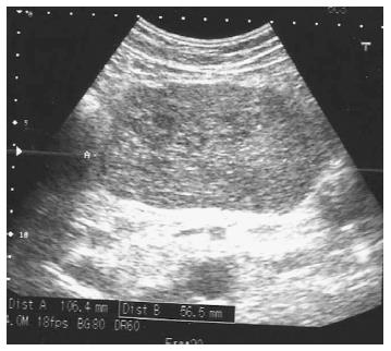
Figure 2 Abdominal ultrasound disclosed hypoechoic, heterogeneous mass filling the epigastrium.
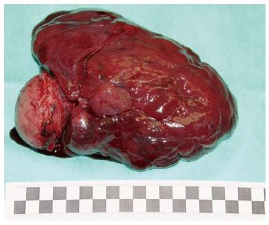
Figure 3 Removed gastric gastrointestinal stromal tumour.
The capsule was not damaged.
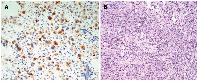
Figure 4 Pathology images.
A: Weak immunoreactivity to KIT (CD 117; original magnification × 200); B: Gastric gastrointestinal stromal tumour (HE staining; original magnification × 100).
- Citation: Lech G, Korcz W, Kowalczyk E, Guzel T, Radoch M, Krasnodębski IW. Giant gastrointestinal stromal tumour of rare sarcomatoid epithelioid subtype: Case study and literature review. World J Gastroenterol 2015; 21(11): 3388-3393
- URL: https://www.wjgnet.com/1007-9327/full/v21/i11/3388.htm
- DOI: https://dx.doi.org/10.3748/wjg.v21.i11.3388


