Copyright
©The Author(s) 2015.
World J Gastroenterol. Mar 21, 2015; 21(11): 3376-3379
Published online Mar 21, 2015. doi: 10.3748/wjg.v21.i11.3376
Published online Mar 21, 2015. doi: 10.3748/wjg.v21.i11.3376
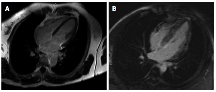
Figure 1 In the “normal” image (A) and “abnormal” image (B) in the late phase of gadolinium enhanced study.
A: No enhancement in noted in the myocardium; B: There is diffuse, heterogeneous enhancement of the left and right ventricular myocardium as well as the interventricular septum in the late phase of gadolinium enhancement study.
- Citation: Fleming K, Ashcroft A, Alexakis C, Tzias D, Groves C, Poullis A. Proposed case of mesalazine-induced cardiomyopathy in severe ulcerative colitis. World J Gastroenterol 2015; 21(11): 3376-3379
- URL: https://www.wjgnet.com/1007-9327/full/v21/i11/3376.htm
- DOI: https://dx.doi.org/10.3748/wjg.v21.i11.3376


