Copyright
©The Author(s) 2015.
World J Gastroenterol. Mar 21, 2015; 21(11): 3367-3375
Published online Mar 21, 2015. doi: 10.3748/wjg.v21.i11.3367
Published online Mar 21, 2015. doi: 10.3748/wjg.v21.i11.3367
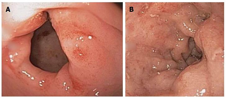
Figure 1 Endoscopic images of gastric antrum and duodenal bulb erosions (A and B).
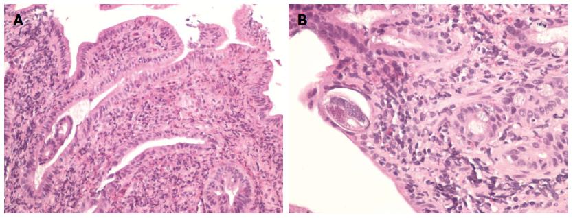
Figure 2 Hematoxylin and Eosin staining of the biopsy specimen.
A: Hematoxylin and eosin (HE) stained section of small bowel biopsy showing prominent eosinophilia associated with Strongyloides infection; B: HE stained longitudinal cross section of a small bowel biopsy showing Strongyloides worm lying within the crypt.
- Citation: Makker J, Balar B, Niazi M, Daniel M. Strongyloidiasis: A case with acute pancreatitis and a literature review. World J Gastroenterol 2015; 21(11): 3367-3375
- URL: https://www.wjgnet.com/1007-9327/full/v21/i11/3367.htm
- DOI: https://dx.doi.org/10.3748/wjg.v21.i11.3367


