Copyright
©The Author(s) 2015.
World J Gastroenterol. Mar 14, 2015; 21(10): 3132-3138
Published online Mar 14, 2015. doi: 10.3748/wjg.v21.i10.3132
Published online Mar 14, 2015. doi: 10.3748/wjg.v21.i10.3132
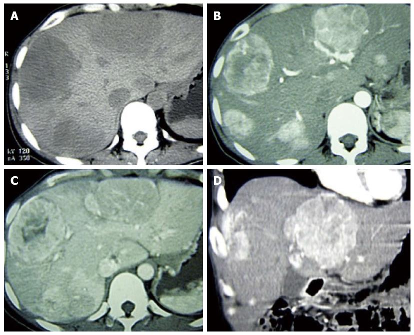
Figure 1 Multidetector computed tomography, axial images.
A: Multiple well-circumscribed, heterogeneous, and hypodense liver masses were present; the largest was located in the right lobe and measured 6.4 cm × 6.3 cm × 5.0 cm; All lesions demonstrated significant enhancement in the (B) arterial and (C) portal phases, with no enhancement of the irregular necrotic area within the largest lesion; D: Several nonenhanced foci were present in other lesions, and a definite contrast washout pattern with peripheral rim enhancement was present in the delayed phase. The background liver tissue was not cirrhotic.
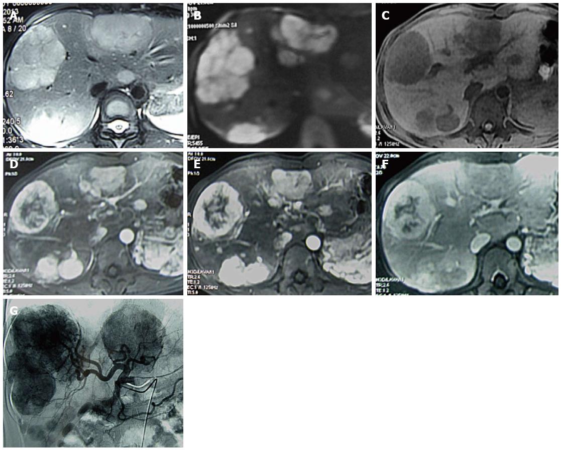
Figure 2 Magnetic resonance images.
A: T2-weighted axial image; and B: diffusion image (b factor = 800) demonstrated well-circumscribed masses of high signal intensity as well as lobulated, irregular masses with low signal intensity; C: Precontrast T1-weighted image showed well-circumscribed, heterogeneous, and hypointense lesions. On enhanced magnetic resonance images, the solid portions of these tumors were significantly enhanced in the early arterial phase; D: irregular to no enhancement was present in the central or foci embellishment of the lesions; E: The enhancement continued in the portal venous phase; F: A definite defect was present in the 20-min delayed hepatobiliary phase find a well-rim arc-shaped artery vessels accompanied on the lesion side on the coronal image; G: Digital subtraction angiography showed multiple hypervascular tumor-staining lesions with sharp edges in the arterial phase.
Figure 3 Microscopic findings of the primary hepatic neuroendocrine tumor in the present case.
A: Tumor cells surrounded a glass-like substance exhibiting a cylindrical arrangement of red staining cytoplasm, and the nucleus was darkly stained (high-power field, × 200); B and C: Immunohistochemical synaptophysin and pancreatic and duodenal homeobox 1 staining in positive tumor cells, respectively.
- Citation: Yang K, Cheng YS, Yang JJ, Jiang X, Guo JX. Primary hepatic neuroendocrine tumor with multiple liver metastases: A case report with review of the literature. World J Gastroenterol 2015; 21(10): 3132-3138
- URL: https://www.wjgnet.com/1007-9327/full/v21/i10/3132.htm
- DOI: https://dx.doi.org/10.3748/wjg.v21.i10.3132


