Copyright
©2014 Baishideng Publishing Group Co.
World J Gastroenterol. Feb 7, 2014; 20(5): 1318-1324
Published online Feb 7, 2014. doi: 10.3748/wjg.v20.i5.1318
Published online Feb 7, 2014. doi: 10.3748/wjg.v20.i5.1318
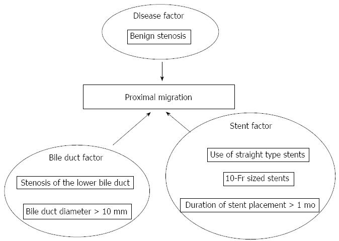
Figure 1 Potential risk factors.
The potential risk factors for migration included bile duct stenosis secondary to benign disease (P = 0.030); stenosis of the lower bile duct (P = 0.031); bile duct diameter > 10 mm (P = 0.023); duration of stent placement > 1 mo (P = 0.007); use of straight-type stents (P < 0.001); and 10-Fr stents (P < 0.001).
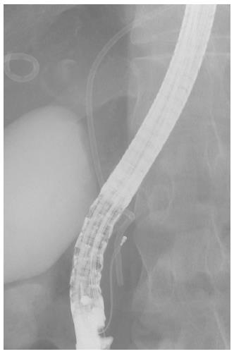
Figure 2 Grasping technique (Case 1).
Examination was performed for hilus bile duct tumor. Pig-tailed-type 7-Fr stent migrated upon stent insertion. An opened eight-wired basket grasped the distal part of the stent. Stent retrieval was successful in Case 1.
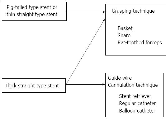
Figure 3 Retrieval methods for migrated stents.
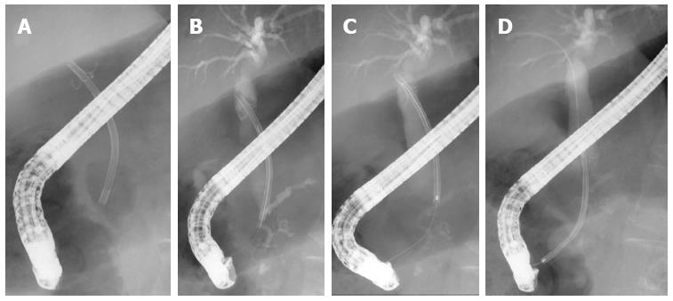
Figure 4 Cannulation technique (Case 2).
A: Biliary drainage was performed for bile duct stenosis due to alcoholic chronic pancreatitis. Straight-type, 10-Fr stent migrated 6 mo after insertion; B: The guidewire was passed through the lumen of the migrated stent; the distal end of the migrated stent was connected with a cannula; C: Migrated stent was pulled, orienting the axis carefully; D: Stent retrieval was successful in Case 2.
- Citation: Kawaguchi Y, Ogawa M, Kawashima Y, Mizukami H, Maruno A, Ito H, Mine T. Risk factors for proximal migration of biliary tube stents. World J Gastroenterol 2014; 20(5): 1318-1324
- URL: https://www.wjgnet.com/1007-9327/full/v20/i5/1318.htm
- DOI: https://dx.doi.org/10.3748/wjg.v20.i5.1318


