Copyright
©2014 Baishideng Publishing Group Co.
World J Gastroenterol. Feb 7, 2014; 20(5): 1180-1191
Published online Feb 7, 2014. doi: 10.3748/wjg.v20.i5.1180
Published online Feb 7, 2014. doi: 10.3748/wjg.v20.i5.1180
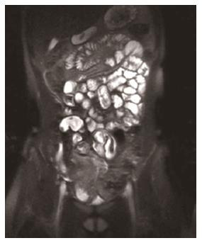
Figure 1 Hydro-magnetic resonance imaging using mannitol orally.
Sometimes the filling result is inadequate and the distension of the bowel is insufficient.
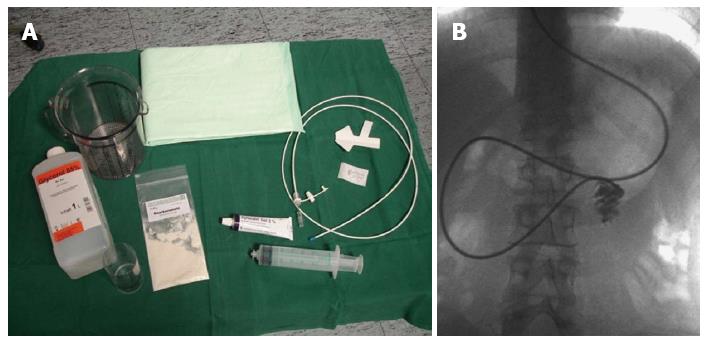
Figure 2 Magnetic resonance enteroclysma requires application of a nasojejunal tube (A) and placement will be guided by pulsed fluoroscopy (B).
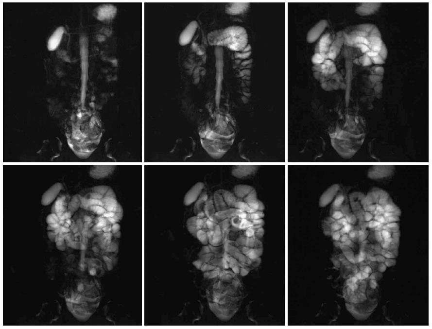
Figure 3 Enteral filling can be followed by using thick slab T2 weighted sequence (7-10 cm thickness).
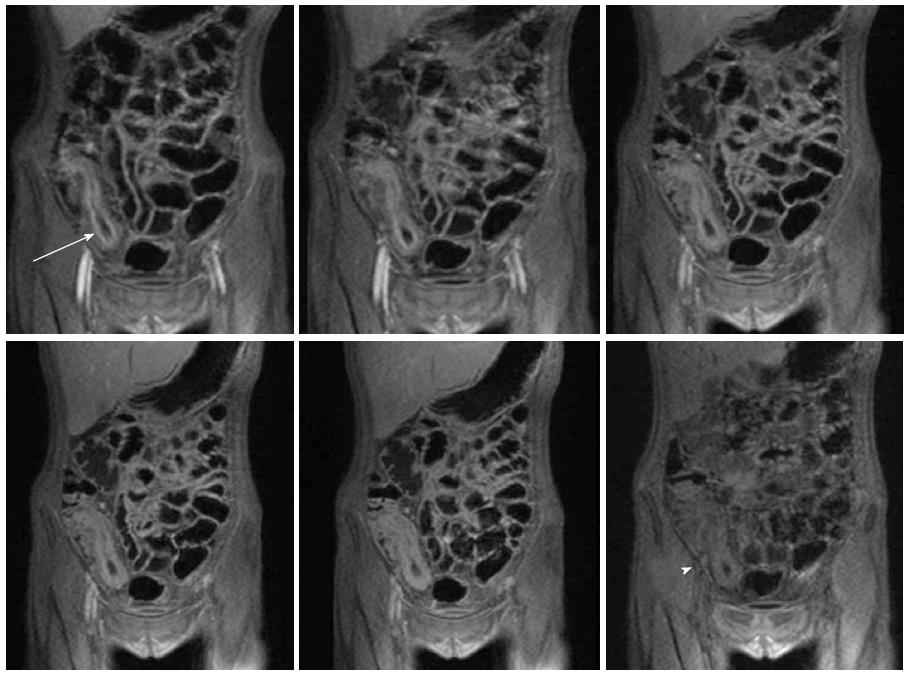
Figure 4 Dynamic contrast enhanced T1-volume interpolated gradient-echo sequence demonstrating different phases of signal increase in the various layers of the bowel wall [at first mucosa (arrow), followed by the serosa, the muscularis, and finally the submucosa (arrowhead)].
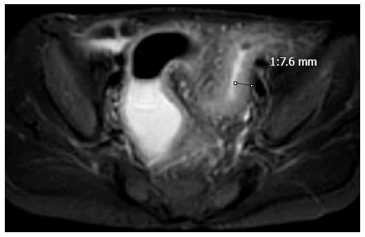
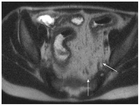
Figure 5 Eleven year old boy with Crohn’s disease.
T2 weighted sequence with fat saturation (Tirm/Stir) demonstrates thickening of the bowel wall and hyperintense signal corresponding to edema in acute inflammation. Narrowing of the lumen.
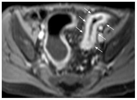
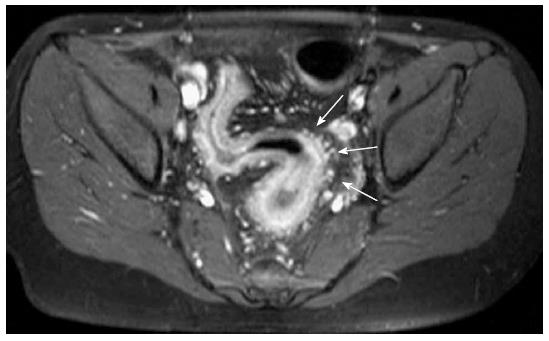
Figure 6 Eleven year old boy with Crohn’s disease.
T1 weighted contrast enhanced sequence shows strong transmural enhancement (arrows).
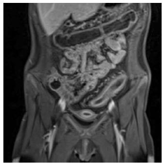
Figure 7 Stratified enhancement in the sigmoid in a female suffering from ulcerative colitis.
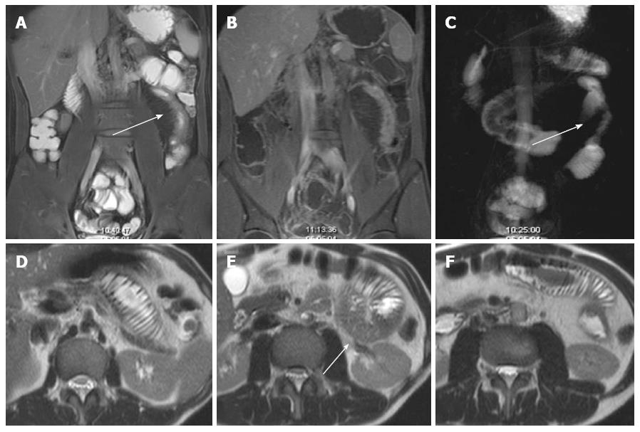
Figure 8 Stricture in inflammatory bowel diseases-chronic phase, no activity of inflammation.
There is no edema with reduced signal intensity in T2 weighted sequences (A, D, arrow). The MR enteroclysma does not show any widening of the involved jejuna bowel loop (C, arrow), also seen in axial T2w sequence (D-F). There is moderate enhancement (B).
Figure 9 Twelve year old female with Crohn’s disease.
Comb sign. CE-T1 weighted sequence with fat saturation. Blood vessels within the mesentery (arrows).
Figure 10 Eleven year old male with Crohn’s disease.
Creeping fat sign (arrows).
Figure 11 Thirteen years old girl with Crohn’s disease.
A: T2w image demonstrating strong hyperintense ascites. Arrow marks the free ascites; B: T1w sequence after contrast application. Fluid is not as easily detected as in T2w images. T1w: T1 weighted; T2w: T2 weighted.
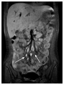
Figure 12 Coronally oriented true steady-state free precession images shows enlarged mesenteric lymph nodes (arrows).
Figure 13 Seventeen years female with Crohn’s disease.
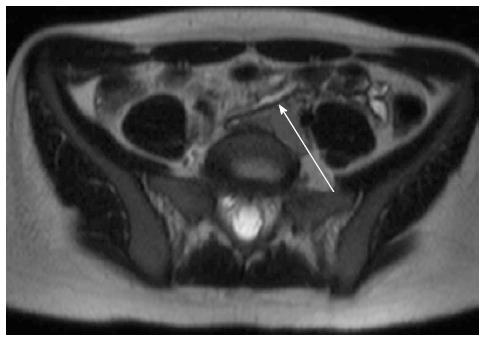
T2 weighted sequence showing enterocolic fistula (arrow).
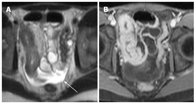
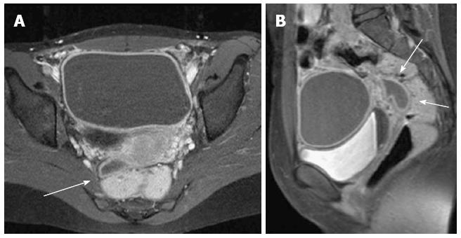
Figure 14 Female with Crohn’s disease.
Sagittal T1 weighted sequence allows to describe presacral abscess (arrow). Fat saturation was applied. A: Axial image; B: Sagittal image (arrows marking abscess; late contrast phase. Notice sedimentation of different components in the bladder).
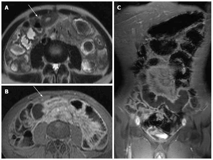
Figure 15 Magnetic resonance enterography allows an overview about involved bowel segments.
In this case there was an isolated involvement of the jejunum. A: Axial T2w sequence showing thickened bowel wall without edema; B, C: Axial and coronal fat saturated contrast-enhanced T1w sequence demonstrating transmural enhancement in Crohn’s disease. T1w: T1 weighted; T2w: T2 weighted.
- Citation: Mentzel HJ, Reinsch S, Kurzai M, Stenzel M. Magnetic resonance imaging in children and adolescents with chronic inflammatory bowel disease. World J Gastroenterol 2014; 20(5): 1180-1191
- URL: https://www.wjgnet.com/1007-9327/full/v20/i5/1180.htm
- DOI: https://dx.doi.org/10.3748/wjg.v20.i5.1180


