Copyright
©2014 Baishideng Publishing Group Inc.
World J Gastroenterol. Dec 21, 2014; 20(47): 17955-17961
Published online Dec 21, 2014. doi: 10.3748/wjg.v20.i47.17955
Published online Dec 21, 2014. doi: 10.3748/wjg.v20.i47.17955
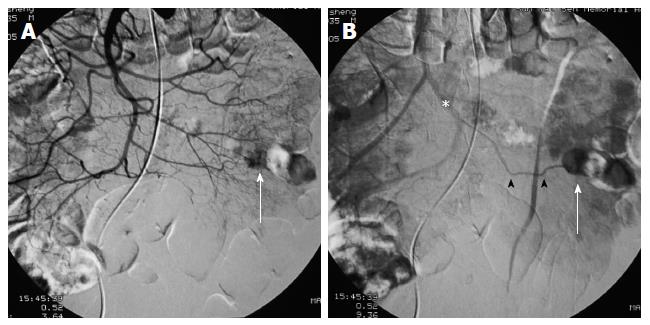
Figure 1 Small bowel gastrointestinal stromal tumor.
A 70-year-old man with a 1.2 cm gastrointestinal stromal tumor of very low risk classification in the upper jejunum. The tumor (white arrows) was round, well-defined and homogeneous in the early arterial phase (A, B). The feeding arteries were not significantly enlarged, and the draining veins (B: black arrowheads) developed clearly and early, merging into the portal vein system (B, asterisk).
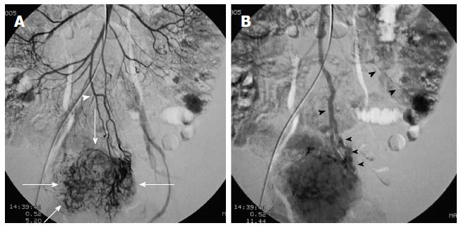
Figure 2 Larger and multiple small bowel gastrointestinal stromal tumors.
A 52-year-old woman with 2 gastrointestinal stromal tumors in the middle jejunum. The tumors (white arrows) were 5.5 cm and 1.5 cm in diameter, respectively, and were also hypervascular and homogeneous in the early arterial phrase. The feeding artery of the large tumor (A: white arrowhead) was slightly thickened. The draining veins of the tumors (B: black arrowheads) were also clear. The pathological risk classification was high (large) and very low (small).
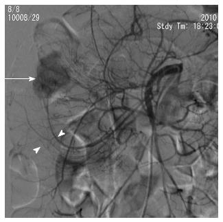
Figure 3 Small bowel gastrointestinal stromal tumor case combined with intussusception.
A 34-year-old woman with tarry stool and abdominal pain. Digital subtraction angiography showed a 2.5 cm well-defined hypervascular tumor (white arrow) in the middle jejunum. The left branches of the superior mesenteric artery shifted to the upper right and was arm-shaped (white arrowheads; intussusception proved by surgery) around the tumor. The pathological risk of this patient was low.
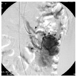
Figure 4 Small bowel gastrointestinal stromal tumor case with active bleeding.
A 35-year-old woman with a 3.2 cm gastrointestinal stromal tumor in the middle jejunum. The hemoglobin was 34 g/L. Digital subtraction angiography showed many draining veins (arrows) on the tumor merging into the portal vein system (arrowheads) without signs of active bleeding. The tumor was slightly lobular and was low risk based on the pathological classification.
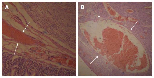
Figure 5 Representative findings from pathological examination of small bowel gastrointestinal stromal tumor.
Two microscopic fields (A, B) of a hematoxylin-eosin stained gastrointestinal stromal tumor (GIST) shows a rich variety of tumor vessels filled with red blood cells (white arrows) (magnification × 40).
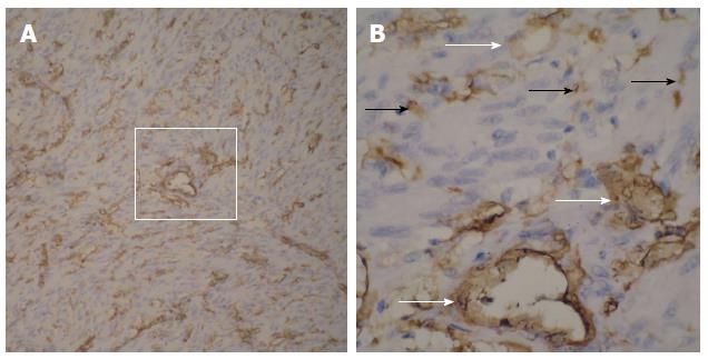
Figure 6 Representative findings from immunohistochemical examination of small bowel gastrointestinal stromal tumor.
A: CD34-positive cells, as well as numerous blood vessels of various diameters (magnification × 100), are shown; B: The magnified imaging (A, white box) in which the CD34-positive tumor cells (B, black arrows) and vascular endothelial cells (B, white arrows) are clearly evident (magnification × 400).
- Citation: Chen YT, Sun HL, Luo JH, Ni JY, Chen D, Jiang XY, Zhou JX, Xu LF. Interventional digital subtraction angiography for small bowel gastrointestinal stromal tumors with bleeding. World J Gastroenterol 2014; 20(47): 17955-17961
- URL: https://www.wjgnet.com/1007-9327/full/v20/i47/17955.htm
- DOI: https://dx.doi.org/10.3748/wjg.v20.i47.17955


