Copyright
©2014 Baishideng Publishing Group Inc.
World J Gastroenterol. Dec 7, 2014; 20(45): 17254-17259
Published online Dec 7, 2014. doi: 10.3748/wjg.v20.i45.17254
Published online Dec 7, 2014. doi: 10.3748/wjg.v20.i45.17254
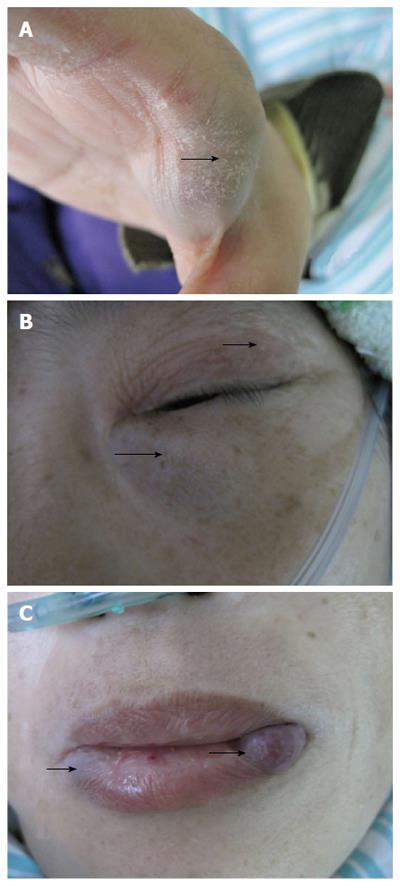
Figure 1 Bluish nodules on upper (A) (arrow) and lower limbs, eyelid (B) (arrows), lip (C) (arrows); vary from 0.
5 to 3.0 cm.
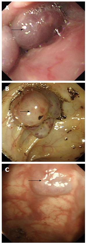
Figure 2 On endoscopy, glottis and esophagus (A) showed multiple bluish hemangiomatas (arrow), and no bleeding was seen; one lesion (2.
0 cm × 2.5 cm) with no fresh bleeding was seen in the colon (B) (arrow), and lesions were also observed on anus (C) (arrow).
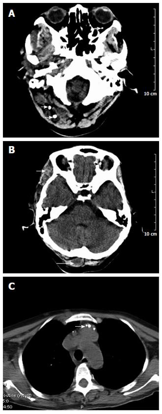
Figure 3 Computed tomography images.
Head computed tomography (CT) (A, B) showed lesions (arrows) on the scalp, left tempora, and orbit, the brain was normal. Chest CT (C) of the root of the neck, clavicle area and superior mediastinum showed multiple nodular, lumpish lesions, and a soft tissue (arrow) component that was consistent with a vascular malformation and hemic calculus. L: Left; P: Posterior.
- Citation: Jin XL, Wang ZH, Xiao XB, Huang LS, Zhao XY. Blue rubber bleb nevus syndrome: A case report and literature review. World J Gastroenterol 2014; 20(45): 17254-17259
- URL: https://www.wjgnet.com/1007-9327/full/v20/i45/17254.htm
- DOI: https://dx.doi.org/10.3748/wjg.v20.i45.17254


